

Characterization of human follicular thyroid cancer cell lines in preclinical mouse models
- PMID: 26830329
- PMCID: PMC5002955
- DOI: 10.1530/EC-15-0114
Characterization of human follicular thyroid cancer cell lines in preclinical mouse models
Abstract
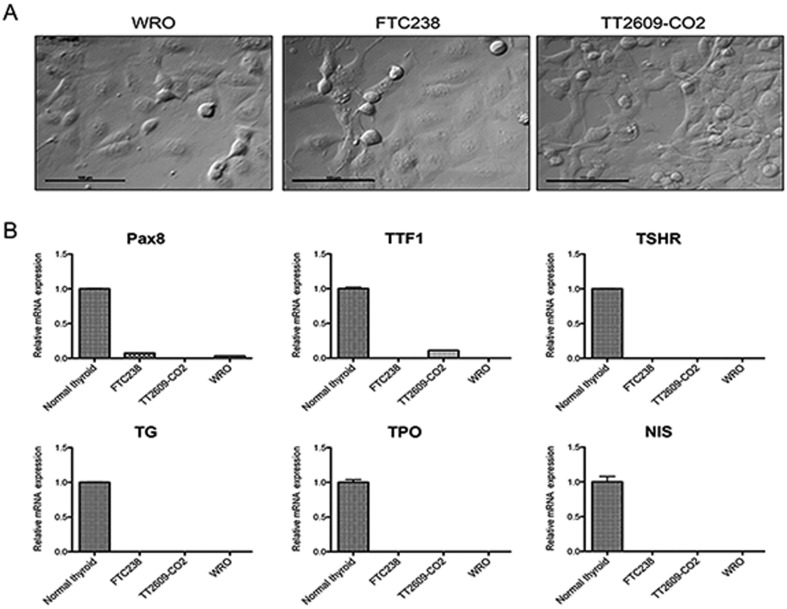
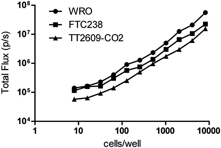
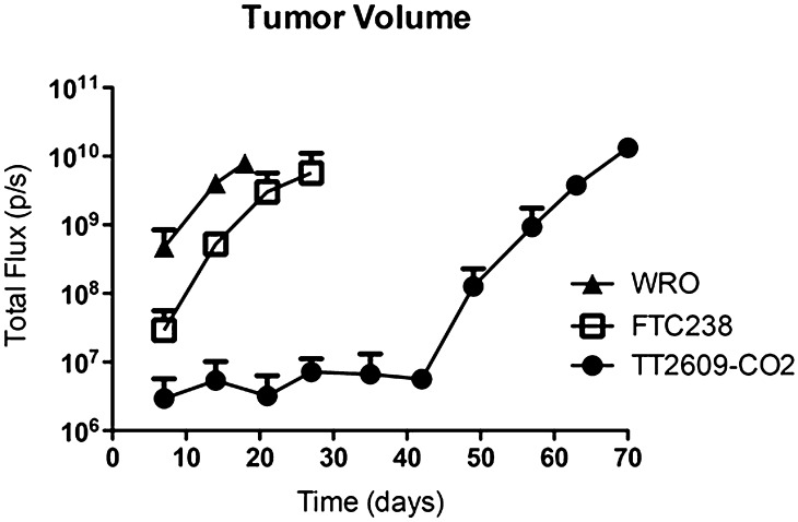
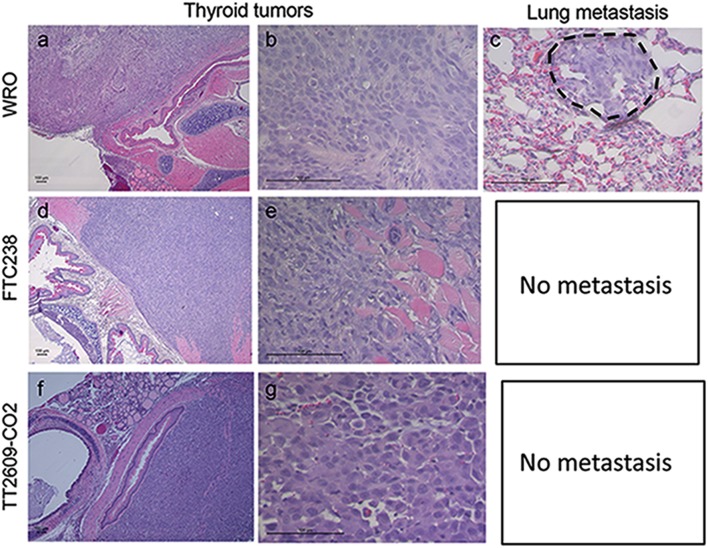
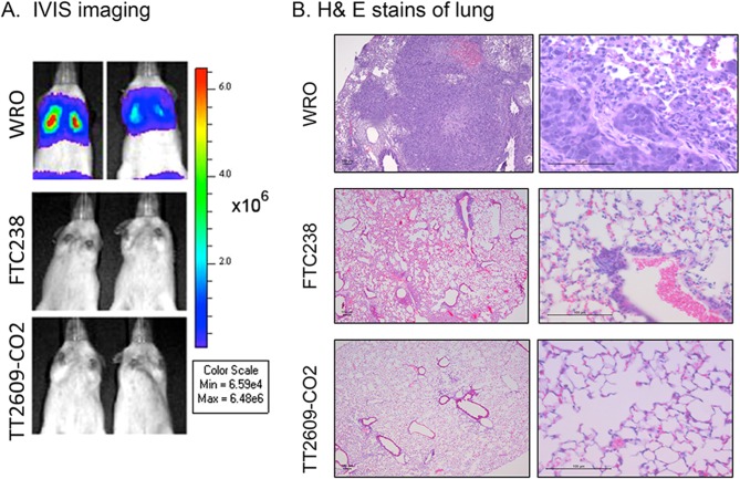
Follicular thyroid cancer (FTC) is the second most common type of thyroid cancers. In order to develop more effective personalized therapies, it is necessary to thoroughly evaluate patient-derived cell lines in in vivo preclinical models before using them to test new, targeted therapies. This study evaluates the tumorigenic and metastatic potential of a panel of three human FTC cell lines (WRO, FTC-238, and TT1609-CO2) with defined genetic mutations in two in vivo murine models: an orthotopic thyroid cancer model to study tumor progression and a tail vein injection model to study metastasis. All cell lines developed tumors in the orthotopic model, with take rates of 100%. Notably, WRO-derived tumors grew two to four times faster than tumors arising from the FTC-238 and TT2609-CO2 cell lines. These results mirrored those of a tail vein injection model for lung metastasis: one hundred percent of mice injected with WRO cells in the tail vein exhibited aggressive growth of bilateral lung metastases within 35 days. In contrast, tail vein injection of FTC-238 or TT2609-CO2 cells did not result in lung metastasis. Together, our work demonstrates that these human FTC cell lines display highly varied tumorigenic and metastatic potential in vivo with WRO being the most aggressive cell line in both orthotopic and lung metastasis models. This information will be valuable when selecting cell lines for preclinical drug testing.
Keywords: follicular thyroid cancer; metastasis; mouse model; orthotopic; thyroid.
© 2016 The authors.
Figures

Similar articles
-
An in vivo mouse model of metastatic human thyroid cancer.Thyroid. 2014 Apr;24(4):695-704. doi: 10.1089/thy.2013.0149. Epub 2014 Mar 10. Thyroid. 2014. PMID: 24262022 Free PMC article.
-
T24 HRAS transformed NIH/3T3 mouse cells (GhrasT-NIH/3T3) in serial tumorigenic in vitro/in vivo passages give rise to increasingly aggressive tumorigenic cell lines T1-A and T2-A and metastatic cell lines T3-HA and T4-PA.Exp Cell Res. 2016 Jan 1;340(1):1-11. doi: 10.1016/j.yexcr.2015.07.029. Epub 2015 Aug 5. Exp Cell Res. 2016. PMID: 26254261
-
Epidermal growth factor enhances proliferation, migration, and invasion of follicular and papillary thyroid cancer in vitro and in vivo.J Clin Endocrinol Metab. 1994 Aug;79(2):401-8. doi: 10.1210/jcem.79.2.8045955. J Clin Endocrinol Metab. 1994. PMID: 8045955
-
On the development of models in mice of advanced visceral metastatic disease for anti-cancer drug testing.Cancer Metastasis Rev. 2007 Dec;26(3-4):737-47. doi: 10.1007/s10555-007-9087-6. Cancer Metastasis Rev. 2007. PMID: 17846863 Review.
-
Animal models of cancer metastasis to the bone.Front Oncol. 2023 Apr 5;13:1165380. doi: 10.3389/fonc.2023.1165380. eCollection 2023. Front Oncol. 2023. PMID: 37091152 Free PMC article. Review.
Cited by
-
Downregulation of miR-146b-3p Inhibits Proliferation and Migration and Modulates the Expression and Location of Sodium/Iodide Symporter in Dedifferentiated Thyroid Cancer by Potentially Targeting MUC20.Front Oncol. 2021 Jan 8;10:566365. doi: 10.3389/fonc.2020.566365. eCollection 2020. Front Oncol. 2021. PMID: 33489878 Free PMC article.
-
Preclinical Imaging for the Study of Mouse Models of Thyroid Cancer.Int J Mol Sci. 2017 Dec 16;18(12):2731. doi: 10.3390/ijms18122731. Int J Mol Sci. 2017. PMID: 29258188 Free PMC article. Review.
-
TERT promoter mutation determines apoptotic and therapeutic responses of BRAF-mutant cancers to BRAF and MEK inhibitors: Achilles Heel.Proc Natl Acad Sci U S A. 2020 Jul 7;117(27):15846-15851. doi: 10.1073/pnas.2004707117. Epub 2020 Jun 19. Proc Natl Acad Sci U S A. 2020. PMID: 32561648 Free PMC article.
-
Piperlongumine Induces Cellular Apoptosis and Autophagy via the ROS/Akt Signaling Pathway in Human Follicular Thyroid Cancer Cells.Int J Mol Sci. 2023 Apr 28;24(9):8048. doi: 10.3390/ijms24098048. Int J Mol Sci. 2023. PMID: 37175755 Free PMC article.
References
-
- Hatakeyama S, Yamamoto H, Ohyama C. Tumor formation assays. Methods in Enzymology 2010. 479 397–411. - PubMed
LinkOut - more resources
Full Text Sources
Other Literature Sources
