Review
. 2016 Jun 1;594(11):2849-66.
doi: 10.1113/JP271139. Epub 2016 Feb 24.

Inositol 1,4,5-trisphosphate receptors and their protein partners as signalling hubs

Affiliations
Review

Inositol 1,4,5-trisphosphate receptors and their protein partners as signalling hubs

David L Prole et al. J Physiol. .

Abstract

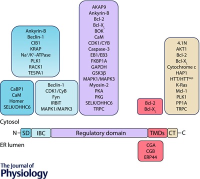
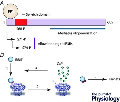
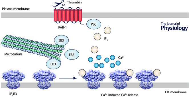
Inositol 1,4,5-trisphosphate receptors (IP3 Rs) are expressed in nearly all animal cells, where they mediate the release of Ca(2+) from intracellular stores. The complex spatial and temporal organization of the ensuing intracellular Ca(2+) signals allows selective regulation of diverse physiological responses. Interactions of IP3 Rs with other proteins contribute to the specificity and speed of Ca(2+) signalling pathways, and to their capacity to integrate information from other signalling pathways. In this review, we provide a comprehensive survey of the proteins proposed to interact with IP3 Rs and the functional effects that these interactions produce. Interacting proteins can determine the activity of IP3 Rs, facilitate their regulation by multiple signalling pathways and direct the Ca(2+) that they release to specific targets. We suggest that IP3 Rs function as signalling hubs through which diverse inputs are processed and then emerge as cytosolic Ca(2+) signals.


Figures

Figure 1
Figure 1. Association of proteins with IP3Rs
Key functional domains of a single IP3R subunit are shown: the suppressor domain (SD), IP3‐binding core (IBC), cytosolic regulatory domain, transmembrane domains (TMDs) and the cytosolic C‐terminus (CT). The sites to which proteins are proposed to bind are shown. Many additional proteins are thought to associate with IP3Rs, but the binding sites have not been identified. Abbreviations and references are provided in Tables 1, 2, 3, 4.
Figure 2
Figure 2. IRBIT controls the sensitivity of IP3Rs
A, the N‐terminal region of IRBIT includes a serine‐rich domain. Phosphorylation of S68, the ‘master’ phosphorylation site, allows sequential phosphorylation of the two residues, S71 and S74, that must be phosphorylated for IRBIT to bind to IP3Rs. Protein phosphatase 1 (PP1) bound to IRBIT dephosphorylates S68. B, phosphorylation of IRBIT (1) allows it to bind to the IBC and so compete with IP3 for binding to the IP3R. Phospho‐IRBIT thereby sets the sensitivity of the IP3R to IP3. IP3 binding to the IBC (2) prevents IRBIT binding and initiates activation of the IP3R. The displaced phospho‐IRBIT can regulate many additional targets, including ion channels and transporters (3). The Ca2+ released by active IP3Rs may control the phosphorylation state of IRBIT, and thereby complete a feedback loop that regulates IP3R sensitivity (4).
Figure 3
Figure 3. A signalling complex assembled around IP3Rs controls gluconeogenesis
Glucagon and insulin exert opposing effects on hepatic gluconeogenesis. Their signalling pathways converge to a protein complex assembled around IP3Rs, the activity of which controls phosphorylation of the transcription factor CRTC2. Dephosphorylated CRTC2 translocates to the nucleus, where it associates with CREB and stimulates transcription of genes required for gluconeogenesis. SIK2 phosphorylates CRTC2, while calcineurin dephosphorylates it. Glucagon, via a GPCR, stimulates both PLC and AC. The IP3 produced by PLC stimulates IP3Rs. The cAMP generated by AC stimulates PKA and that promotes dephosphorylation of CRTC2 by phosphorylating both SIK2 (inhibiting its activity) and IP3Rs, sensitizing the latter to IP3. The larger Ca2+ signal then activates calcineurin. Insulin causes activation of AKT1, which phosphorylates IP3Rs and inhibits their activity; it thereby opposes the effects of glucagon and attenuates calcineurin activity. Phosphorylation is indicated by red circles, black arrows denote stimulation and the red arrow denotes inhibition. Abbreviations and further details in the text and tables.
Figure 4
Figure 4. EB3 is required for effective signalling by IP3Rs in endothelial cells
In endothelial cells, EB3 binds to a TxIP motif within the regulatory domain of IP3R3, allowing IP3Rs to associate with the plus‐end of microtubules. Disrupting this interaction prevents clustering of IP3Rs and attenuates the Ca2+ signals evoked by thrombin, which cleaves within the N‐terminus of PAR‐1 and allows it to stimulate PLC. The evidence (Geyer et al. 2015) suggests that the EB3‐mediated interaction of IP3R3 with microtubules is essential for the clustering of IP3Rs that allows the Ca2+ released by one IP3R to be amplified by recruitment of neighbouring IP3Rs.

References

    1. Alzayady KJ, Panning MM, Kelley GG & Wojcikiewicz RJ (2005). Involvement of the p97‐Ufd1‐Npl4 complex in the regulated endoplasmic reticulum‐associated degradation of inositol 1,4,5‐trisphosphate receptors. J Biol Chem 280, 34530–34537. - PMC - PubMed
    1. Ando H, Kawaai K & Mikoshiba K (2014). IRBIT: A regulator of ion channels and ion transporters. Biochim Biophys Acta 1843, 2195–2204. - PubMed
    1. Ando H, Mizutani A, Matsu‐ura T & Mikoshiba K (2003). IRBIT, a novel inositol 1,4,5‐trisphosphate (IP3) receptor‐binding protein, is released from the IP3 receptor upon IP3 binding to the receptor. J Biol Chem 278, 10602–10612. - PubMed
    1. Angrand PO, Segura I, Volkel P, Ghidelli S, Terry R, Brajenovic M, Vintersten K, Klein R, Superti‐Furga G, Drewes G, Kuster B, Bouwmeester T & Acker‐Palmer A (2006). Transgenic mouse proteomics identifies new 14‐3‐3‐associated proteins involved in cytoskeletal rearrangements and cell signaling. Mol Cell Proteomics 5, 2211–2227. - PubMed
    1. Arnaoutov A & Dasso M (2014). Enzyme regulation. IRBIT is a novel regulator of ribonucleotide reductase in higher eukaryotes. Science 345, 1512–1515. - PMC - PubMed

Publication types

MeSH terms

Substances