. 2016 Feb;57(2):320-31.
doi: 10.1167/iovs.15-18716.

Palisade Endings Are a Constant Feature in the Extraocular Muscles of Frontal-Eyed, But Not Lateral-Eyed, Animals

Affiliations

Palisade Endings Are a Constant Feature in the Extraocular Muscles of Frontal-Eyed, But Not Lateral-Eyed, Animals

Roland Blumer et al. Invest Ophthalmol Vis Sci. 2016 Feb.

Abstract

Purpose: To test whether palisade endings are a general feature of mammalian extraocular muscles (EOMs).

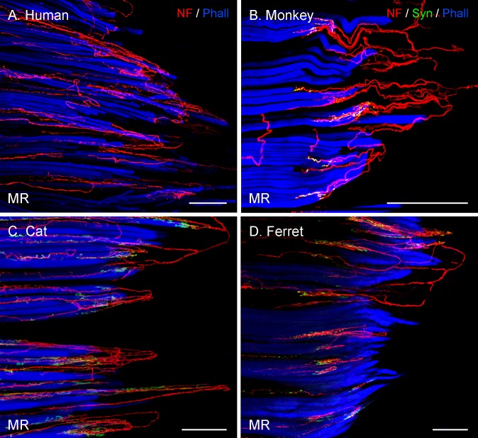
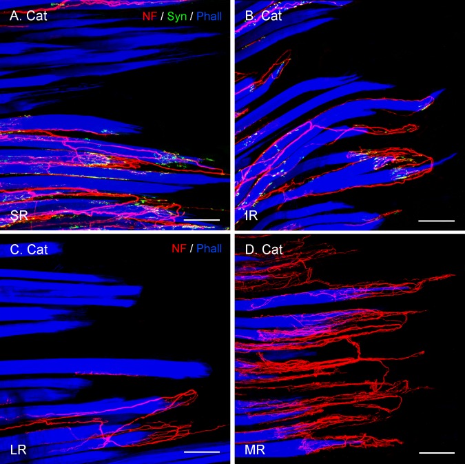
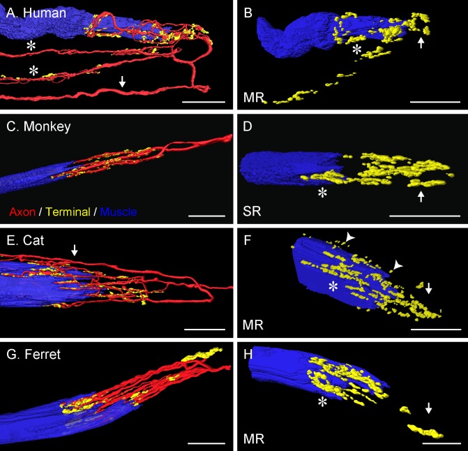
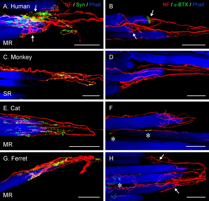
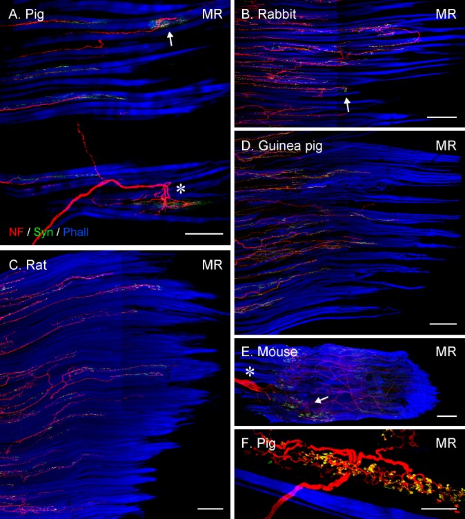
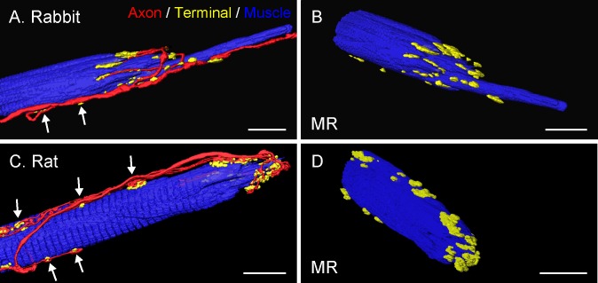
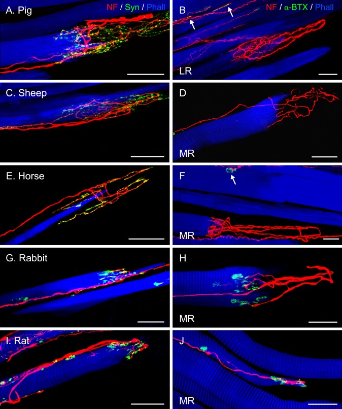
Methods: Thirteen species, some frontal-eyed (human, monkey, cat, and ferret), and others lateral-eyed (pig, sheep, calf, horse, rabbit, rat, mouse, gerbil, and guinea pig) were analyzed. Palisade endings were labeled by using different combinations of immunofluorescence techniques. Three-dimensional reconstructions of immunolabeled palisade endings were done.

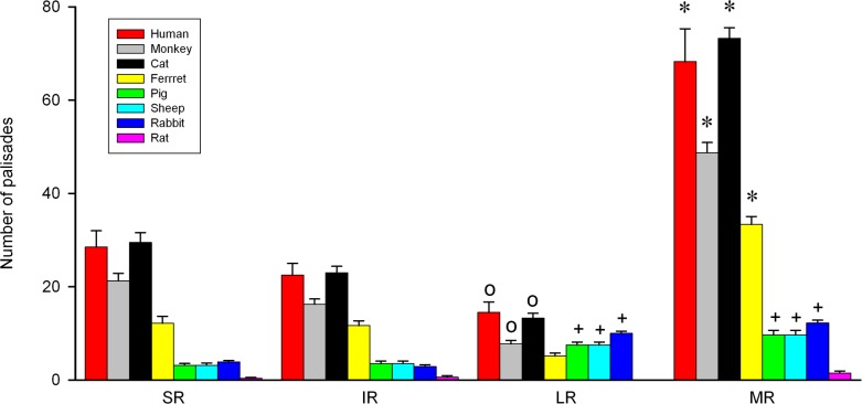
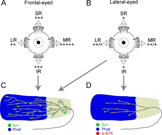
Results: In all frontal-eyed species, palisade endings were a consistent feature in the rectus EOMs. Their total number was high and they exhibited an EOM-specific distribution. In particular, the number of palisade endings in the medial recti was significantly higher than in the other rectus muscles. In the lateral-eyed animals, palisade endings were infrequent and, when present, their total number was rather low. They were only found in ungulates (sheep, calf, pig, and horse) and in rabbit. In rodents (rat, guinea pig, mouse, and gerbil) palisade endings were found infrequently (e.g., rat) or were completely absent. Palisade endings in frontal-eyed species and in some lateral-eyed species (pig, sheep, calf, and horse) had a uniform morphology. They generally lacked α-bungarotoxin staining, with a few exceptions in primates. Palisade endings in other lateral-eyed species (rabbit and rat) exhibited a simplified morphology and bound α-bungarotoxin.

Conclusions: Palisade endings are not a universal feature of mammalian EOMs. So, if they are proprioceptors, not all species require them. Because in frontal-eyed species, the medial rectus muscle has the highest number of palisade endings, they likely play a special role in convergence.


Figures

Figure 1
Figure 1
Bar chart showing the total number and the muscle-specific distribution of palisade endings. The total number of palisade endings was higher in frontal-eyed than lateral-eyed species. In frontal-eyed species, the medial rectus muscle (MR) always contained the highest number of palisade endings and the lateral rectus (LR) the lowest. The values of the vertical eye muscles (superior rectus, SR, and inferior rectus, IR) were similar to each other. In lateral-eyed species, the MR and LR had more palisade endings than the SR and IR, except in the rat where the number of palisade endings was extremely low. Data represent mean and SEM. Eight muscles for each EOM were analyzed in cat, rabbit, and rat; six muscles in ferret, pig, and sheep; four in monkey and human LR and MR; and two for human SR and IR. *Significantly higher number of palisade endings in MR with respect to the other muscles. +, significant differences of MR and LR with SR and IR; O, significantly lower number of palisade endings in LR than in the other three muscles (2-way ANOVA test followed by Holm-Sidak method for post hoc multiple comparisons at a significance level of 0.05).
Figure 2
Figure 2
Projection of CLSM z-stacks showing the distal muscle–tendon junction of MR muscles in frontal-eyed species. Only segments of the muscle–tendon junction are shown due to the size of the muscle. (A) Axons were labeled with anti-neurofilament (NF, red) and muscle fibers with phalloidin (Phall, blue). (B, D) Additional labeling of nerve terminals with anti-synaptophysin (Syn, green). In these and other photomicrographs, the tendon extends from the muscles to the right of the image and is not visible. (AD) A high number of palisade endings were detected in the MR. (BD) Terminal branches exhibited synaptophysin-positive varicosities. Scale bars: 200 μm.
Figure 3
Figure 3
Projection of CLSM z-stacks showing segments of the distal muscle–tendon junction of the four rectus muscles (SR, IR, LR, MR) in cat. (A, B) Axons were labeled with anti-neurofilament (NF, red), anti-synaptophysin (Syn, green), and muscle fibers with phalloidin (Phall, blue). (C, D) Staining with anti-neurofilament and phalloidin, but lacking anti-synaptophysin labeling. (AD) The relative EOM-specific abundance of palisade endings with the medial rectus (D) highest and the lateral rectus (C) lowest. The values of the vertical eye muscles (A, B) are in-between. Scale bars: 100 μm.
Figure 4
Figure 4
Three-dimensional reconstruction of palisade endings in frontal-eyed species. In monkey (C), the palisade ending is from an SR muscle; in the other species they are from MR muscles. Nerve fibers were labeled with anti-neurofilament (red), terminal varicosities with anti-synaptophysin (green), and muscle fibers with phalloidin (blue). The yellow color represents the merging of green (synaptophysin) and red (neurofilament) signals. (A, E) Showing the whole formation of the palisade ending with the axon (arrows) indicated. (A) The axon supplies three palisade endings, of which only the upper one is completely shown. Asterisks indicate the collateral supplying the other palisade endings. (C, G) From the axon supplying the palisade endings, only the recurrent part coming from the tendon is shown. (B, D, F, H) Showing the same palisade endings as in (A, C, E, G), respectively, but rotated and the nerve fibers removed. Terminal varicosities are found far away from the muscle fiber (arrows), at the tendon level, and around the muscle fiber tip (asterisks). Many nerve terminals around the muscle fiber tip are separated from the muscle fiber surface by a small gap (arrowheads [F]). Scale bars: 30 μm.
Figure 5
Figure 5
Projection of z-stacks from CLSM images showing palisade endings in frontal-eyed species. All palisade endings are from MR muscles with the exception of (C), which is from an SR muscle. (A, C, E, G) Staining was done with anti-neurofilament (NF, red), anti-synaptophysin (Syn, green), and phalloidin (Phall, blue). (B, D, F, H) Staining with anti-neurofilament, α-bungarotoxin (α-BTX, green), and phalloidin. (A, B, H) Two palisade endings (arrows) in each panel are shown supplying neighboring muscle fibers. In the rest of the images, only a single palisade ending is demonstrated. (A, C, E, G) Terminal varicosities of palisade endings exhibit synaptophysin immunoreactivity. (D, F, H) Absence of α-bungarotoxin signal associated with palisade endings. (F, H) α-Bungarotoxin-positive neuromuscular contacts (asterisks) are on neighboring muscle fibers. (B) Showing two human palisade endings (arrows); the upper one is associated with α-bungarotoxin staining, but not the lower one. Scale bars: 50 μm.
Figure 6
Figure 6
Projection of CLSM z-stacks showing the distal muscle–tendon junction of MR muscles (AD), a whole MR muscle (E) and a Golgi tendon organ (F) in lateral-eyed species. (A) and (B) illustrating only segments of the muscle–tendon junctions, whereas (C) and (D) the entire muscle-tendon junctions. Axons were labeled with anti-neurofilament (NF, red), nerve terminals with anti-synaptophysin (Syn, green), and muscle fibers with phalloidin (Phall, blue). Only in pig (A) and rabbit (B), are individual palisade endings (arrow) seen, and most axons stop at variable distances away from the muscle–tendon junction. Such axons establish numerous synaptophysin-positive contacts, indicating they supply multiply innervated muscle fibers. (A) A Golgi tendon organ (asterisk) is visible as well. In (CE), all axons stop before reaching the muscle–tendon junction and no palisade endings are present. (E) Showing the nerve entry site (asterisk) and the motor endplate zone (arrow) in the proximal part of the muscle. (I) High magnification image of a Golgi tendon organ with synaptophysin-positive nerve terminals. Scale bars: (A, C) 200 μm; (B, D, E) 300 μm; (F) 50 μm.
Figure 7
Figure 7
Three-dimensional reconstruction of palisade endings in lateral-eyed species. All examples are from MR muscles. Nerve fibers were labeled with anti-neurofilament (red), nerve terminals with anti-synaptophysin (green), and muscle fibers with phalloidin (blue). (A, C) The whole extent of the palisade ending is shown with nerve fibers, nerve terminals, and muscle fibers. Axons forming palisade endings establish nerve terminals (arrows) alongside the muscle fibers. The palisade complex itself appears rather simple, without extensive axonal branching and with terminal varicosities around the muscle fiber tip. (B, D) Showing the same palisade endings in different views with the nerve fibers removed. Scale bars: (A, B) 25 μm; (C, D) 15 μm.
Figure 8
Figure 8
Projection of z-stacks from CLSM images showing palisade endings in lateral-eyed species. Palisade endings are from LR muscles in (A, B), and the rest from MR muscles. (A, C, E, G, I) Staining with anti-neurofilament (NF, red), anti-synaptophysin (Syn, green), and phalloidin (Phall, blue), and (B, D, F, H, J) with anti-neurofilament, α-bungarotoxin (α-BTX), and phalloidin. (A, C, E, G, I) Terminal varicosities in palisade endings exhibit synaptophysin immunoreactivity. In pig (A), sheep (C), and horse (E), terminal varicosities of palisade endings are found at the tendon level and also around the muscle fiber tip, whereas in rabbit (G) and rat (I), varicosities of palisade endings are only at the muscle fiber tip. (B, D, F) Alpha-bungarotoxin labeling is generally absent in palisade endings, but is found on neighboring muscle fibers ([B, F] arrows). However, in (H) and (J), α-bungarotoxin is present in association with palisade endings. Scale bars: (AG) 50 μm; (HJ) 25 μm.
Figure 9
Figure 9
Schematic drawing summarizing the distribution pattern and molecular properties of palisade endings in frontal-eyed and lateral-eyed species. In (A) and (B), relative density of palisades is indicated by + in the SR, IR, LR, and MR (differences between rats and nonrodent lateral-eyed species indicated by a ++/+). Frontal-eyed species (A) showed a different specific distribution of palisade endings in the rectus muscles from lateral-eyed species (B). (C) Palisade endings in frontal-eyed species had terminal varicosities at the tendon level and around the muscle fiber tip. Terminal varicosities displayed synaptophysin immunoreactivity (Syn), whereas α-bungarotoxin labeling (α-BTX) was exceptional. (D) In rabbit and rat, palisades exhibited a more simplified morphology and lacked terminal varicosities at the tendon level, but they displayed synaptophysin and α-bungarotoxin staining. However, ungulates exhibited palisade endings whose structure and molecular properties came closer to those found in frontal-eyed animals (C). The tendon in (C) and (D) is illustrated in gray in continuity with the phalloidin (Phall)-stained muscle fiber in blue.

Comment in

Similar articles

Cited by

References

    1. Maier A,, DeSantis M,, Eldred E. The occurrence of muscle spindles in extraocular muscles of various vertebrates. J Morphol. 1974; 143: 397–408. - PubMed
    1. Ruskell GL. The fine structure of human extraocular muscle spindles and their potential proprioceptive capacity. J Anat. 1989; 167: 199–214. - PMC - PubMed
    1. Blumer R,, Lukas JR,, Aigner M,, Bittner R,, Baumgartner I,, Mayr R. Fine structural analysis of extraocular muscle spindles of a two-year-old human infant. Invest Ophthalmol Vis Sci. 1999; 40: 55–64. - PubMed
    1. Greene T,, Jampel R. Muscle spindles in the extraocular muscles of the macaque. J Comp Neurol. 1966; 126: 547–549. - PubMed
    1. Büttner-Ennever JA,, Konakci KZ,, Blumer R. Sensory control of extraocular muscles. Prog Brain Res. 2006; 151: 81–93. - PubMed

Publication types

MeSH terms