Review
. 2016 Mar;10(1):95-108.
doi: 10.1007/s12105-016-0697-6. Epub 2016 Feb 1.

Update on Select Benign Mesenchymal and Meningothelial Sinonasal Tract Lesions

Affiliations
Review

Update on Select Benign Mesenchymal and Meningothelial Sinonasal Tract Lesions

Lester D R Thompson et al. Head Neck Pathol. 2016 Mar.

Abstract

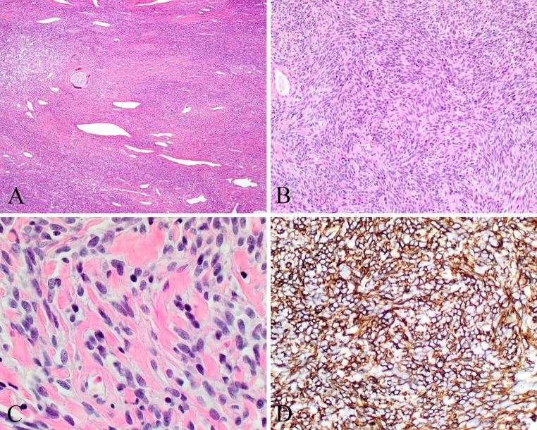
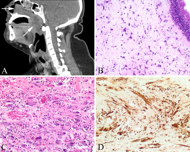
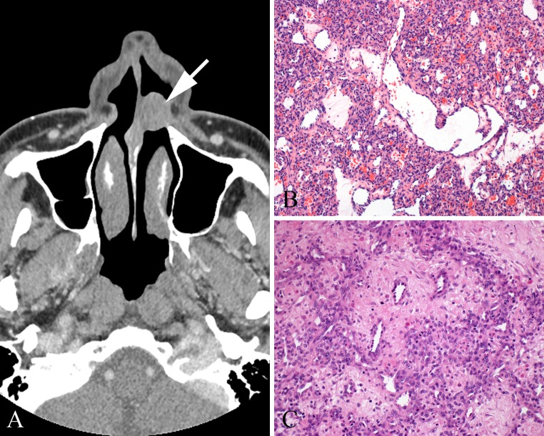
Several benign and malignant mesenchymal and meningothelial lesions may preferentially affect or extend into the sinonasal tract. Glomangiopericytoma (GPC, formerly sinonasal-type hemangiopericytoma) is a specific tumor with a predilection to the sinonasal tract. Sinonasal tract polyps with stromal atypia (antrochoanal polyp) demonstrate unique histologic findings in the sinonasal tract. Juvenile nasopharyngeal angiofibroma (JNA) arises from specialized tissue in this location. Meningioma may develop as direct extension from its intracranial counterpart or as an ectopic tumor. Selected benign mesenchymal tumors may arise in the sinonasal tract and pose a unique differential diagnostic consideration, such as solitary fibrous tumor and GPC or lobular capillary hemangioma and JNA. Although benign and malignant vascular, fibrous, fatty, skeletal muscle, and nerve sheath tumors may occur in this location, this paper focuses on a highly select group of rare benign sinonasal tract tumors with their clinicopathological and molecular findings, and differential diagnosis.

Keywords: Angiofibroma; Diagnosis; Differential; Granuloma; Hemangiopericytoma; Meningioma; Nerve sheath neoplasms; Polyps; Pyogenic; Sinonasal tract; Solitary fibrous tumor.


Figures

Fig. 1
Fig. 1
Glomangiopericytoma (GPC). a Intact surface epithelium overlying a patternless proliferation with well developed peritheliomatous hyalinization. b Oval to elongated nuclei in syncytial cells. Extravasated erythrocytes and eosinophils are noted. c Strong and diffuse smooth muscle actin reaction. d Nuclear β-catenin reaction in the neoplastic cells
Fig. 2
Fig. 2
Solitary fibrous tumor (SFT). a Patulous and open vessels in a cellular fibrous connective tissue stroma. b Haphazard arrangement of spindled tumor cells. c Keloid-like collagen deposition between spindled cells with ovoid nuclei. d Strong and diffuse cytoplasmic-membrane reaction with CD34
Fig. 3
Fig. 3
Antrochoanal polyp (ACP). a A large polyp (arrows) begins in the maxillary sinus and extends into the nasopharynx. b Intact respiratory epithelium with submucosal stellate fibroblasts. c The atypical fibroblastic cells are often identified around areas of vascular injury, associated with hemorrhage. d The fibroblasts show a strong and diffuse reaction with smooth muscle actin
Fig. 4
Fig. 4
Juvenile nasopharyngeal angiofibroma (JNA). a Grossly, a large, fibrous polypoid mass often takes the shape of the nasal cavity. b Variable size and type of vessels in a collagenized stroma. c Smooth muscle walled vessels and large patulous vessels within intervening erectile tissue type collagenized stroma. d Thick, muscled-walled vessels, along with several other vessel types comprise a JNA
Fig. 5
Fig. 5
Lobular capillary hemangioma (LCA). a A septal mass (Little area, arrow) is a characteristic location. b Large central vessel with small ramifying capillaries. This lesion appears cellular because of proliferation of pericytes surrounding proliferated endothelial cells of capillaries; normal form mitotic activity may be abundant. c Lobular arrangement of vessels around a central penetrating vessel
Fig. 6
Fig. 6
Meningioma. a The neoplasm is noted invading between the minor mucoserous glands. b A lobule of tumor cells below an intact respiratory epithelium. c Meningothelial pattern of growth, with whorls. Intranuclear cytoplasmic inclusions are noted. Psammoma bodies are present. d Strong, but focal EMA immunoreactivity is observed

References

    1. Stout AP, Murray MR. Hemangiopericytoma. A vascular tumor featuring Zimmerman’s pericytes. Ann Surg. 1942;116:26–33. doi: 10.1097/00000658-194207000-00004. - DOI - PMC - PubMed
    1. Gold JS, Antonescu CR, Hajdu C, et al. Clinicopathologic correlates of solitary fibrous tumors. Cancer. 2002;94:1057–1068. doi: 10.1002/cncr.10328. - DOI - PubMed
    1. Suster S, Nascimento AG, Miettinen M, et al. Solitary fibrous tumors of soft tissue. A clinicopathologic and immunohistochemical study of 12 cases. Am J Surg Pathol. 1995;19:1257–1266. doi: 10.1097/00000478-199511000-00005. - DOI - PubMed
    1. Miettinen M, Sarlomo-Rikala M, Kovatich AJ. Cell-type- and tumour-type-related patterns of bcl-2 reactivity in mesenchymal cells and soft tissue tumours. Virchows Arch. 1998;433:255–260. doi: 10.1007/s004280050244. - DOI - PubMed
    1. Miettinen M, Paal E, Lasota J, et al. Gastrointestinal glomus tumors: a clinicopathologic, immunohistochemical, and molecular genetic study of 32 cases. Am J Surg Pathol. 2002;26:301–311. doi: 10.1097/00000478-200203000-00003. - DOI - PubMed

LinkOut - more resources