

Review
doi: 10.1007/s12105-016-0690-0.
Epub 2016 Feb 1.
Pathology of Fungal Rhinosinusitis: A Review
Affiliations
- PMID: 26830404
- PMCID: PMC4746136
- DOI: 10.1007/s12105-016-0690-0
Item in Clipboard
Review
Pathology of Fungal Rhinosinusitis: A Review
Head Neck Pathol.
2016 Mar.
Abstract
Fungal rhinosinusitis (FRS) comprises a spectrum of disease processes that vary in clinical presentation, histologic appearances, and biological significance. FRS can be acute or chronic and is most commonly classified as non-invasive or invasive based on whether fungi have invaded into tissue. This manuscript will review the pathologic classification of FRS.
Keywords: Acute invasive fungal rhinosinusitis; Allergic fungal rhinosinusitis; Allergic mucin; Aspergillus; Fungal ball; Rhinosinusitis.
Figures
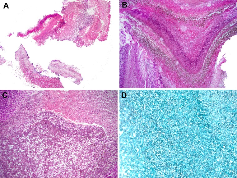
Fungal ball a low power appearance of a maxillary sinus fungal ball from a 60 yo female. Fungal ball is composed of an entangled mass of fungal organisms (Hematoxylin and eosin; original magnification ×10). b Low power of a fungal ball showing a lamellated appearance, which can be confused with eosinophilic (allergic) mucin (EM). This fungal ball shows a layered appearance and pigmented fungal forms. Cultures grew Aspergillus niger (Hematoxylin and eosin; original magnification ×25). c Higher power of fungal ball showing an entangled mass of fungal organisms with very little inflammation (Hematoxylin and eosin; original magnification ×50). d Silver stain highlighting fungal organisms in a fungal ball (Grocott stain; original magnification ×100)
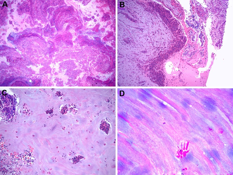
Allergic fungal rhinosinusitis a low power of eosinophilic (allergic) mucin showing a layered appearance of mucin admixed with inflammatory cells and debris (Hematoxylin and eosin; original magnification ×10). b Eosinophilic mucin showing collections of eosinophils and sloughed epithelial cells (Hematoxylin and eosin; original magnification ×50). c Eosinophilic mucin showing eosinophils singly and in cluster (Hematoxylin and eosin; original magnification ×50). d Eosinophilic mucin showing Charcot–Leyden crystals (Hematoxylin and eosin; original magnification ×200)
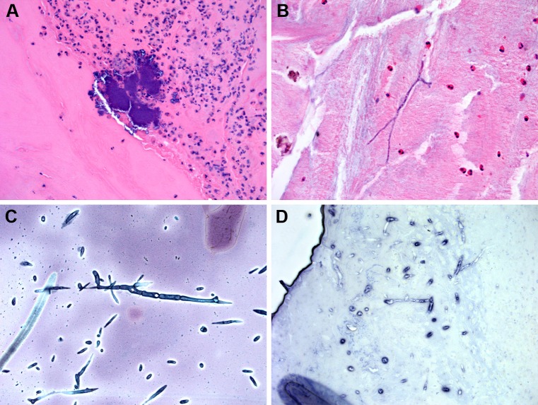
a Eosinophilic mucin often contains bacterial colonies (Hematoxylin and eosin; original magnification ×100). b Rarely, fungal organisms may be seen in eosinophilic mucin without use of specials stains. Cultures grew Curvularia sp. (Hematoxylin and eosin; original magnification ×100). c Silver stain of fungal organisms in eosinophilic mucin. Cultures grew Alternaria alternata (Grocott stain; original magnification ×200). d In situ hybridization for Aspergillus ribosomal RNA in eosinophilic mucin. Cultures confirmed A. fumigatus. (Nitroblue tetrazolium violet; original magnification ×100)
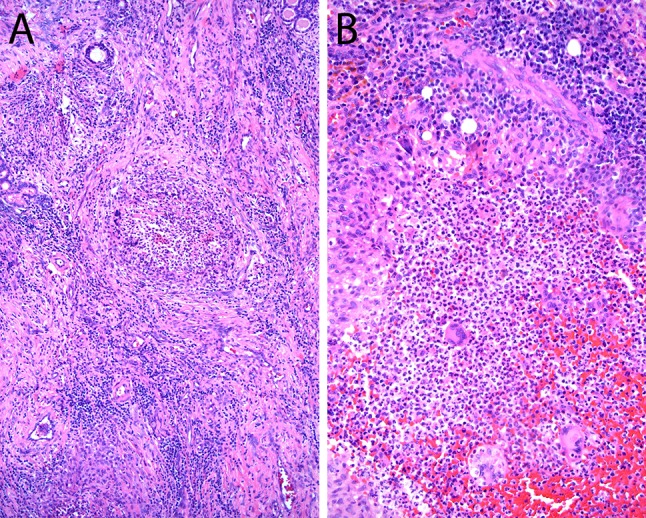
Acute invasive fungal rhinosinusitis a low power view of infarcted sinonasal without significant inflammatory reaction in immunosuppressed patient with acute leukemia with acute invasive fungal rhinosinusitis Culture grew Rhizopus sp. (Hematoxylin and eosin; original magnification ×12.5). b Acute invasive FRS showing fungal organisms invading blood vessels and soft tissue. Cultures grew Aspergillus fumigatus (Hematoxylin and eosin; original magnification ×100). c Silver staining highlighting fungal hyphae in soft tissue in acute invasive FRS. Cultures grew A. fumigatus (Hematoxylin and eosin; original magnification ×100). d In situ hybridization (ISH) for Aspergillus ribosomal RNA. Note the extensive necrosis and only rare positive organisms. rRNA ISH is not always reliable on necrotic tissues (Fast red tetrazolium violet; original magnification ×100)
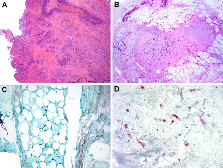
Chronic invasive granulomatous fungal rhinosinusitis a, b Granulomatous reaction toward Aspergillus flavus in patients with chronic invasive granulomatous fungal rhinosinusitis (Hematoxylin and eosin; original magnification ×25 for a and ×100 for b)
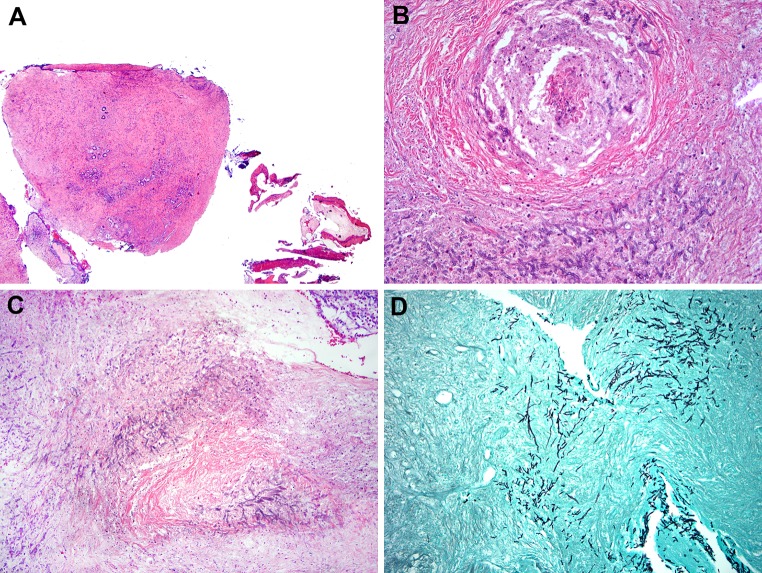
Chronic invasive fungal rhinosinusitis a low power of inflamed and fibrotic sinonasal mucosa in liver transplant patient with chronic invasive fungal rhinosinusitis (Hematoxylin and eosin; original magnification ×12.5). b, c Chronic invasive FRS with extensive sinonasal mucosal necrosis/infarction with visible fungal organisms in liver transplant patient with symptoms greater than 3 months duration. b Fungi within blood vessels (Hematoxylin and eosin; original magnification ×100). d Silver stain shows fungal hyphae infiltrating sinonasal mucosa in chronic invasive FRS (Hematoxylin and eosin; original magnification ×50)
Similar articles
-
Spectrum of fungal rhinosinusitis; histopathologist's perspective.Histopathology. 2009 Jun;54(7):854-9. doi: 10.1111/j.1365-2559.2009.03309.x. Histopathology. 2009. PMID: 19635105
-
'Eosinophilic fungal rhinosinusitis': a common disorder in Europe?Laryngoscope. 2003 Feb;113(2):264-9. doi: 10.1097/00005537-200302000-00013. Laryngoscope. 2003. PMID: 12567080
-
Controversies surrounding the categorization of fungal sinusitis.Med Mycol. 2009;47 Suppl 1:S299-308. doi: 10.1080/13693780802213357. Epub 2008 Jul 28. Med Mycol. 2009. PMID: 18663658 Review.
-
Fungal rhinosinusitis: a categorization and definitional schema addressing current controversies.Laryngoscope. 2009 Sep;119(9):1809-18. doi: 10.1002/lary.20520. Laryngoscope. 2009. PMID: 19544383 Free PMC article. Review.
-
Fungal Rhinosinusitis: An integrated diagnostic approach.Ann Diagn Pathol. 2025 Apr;75:152415. doi: 10.1016/j.anndiagpath.2024.152415. Epub 2024 Nov 26. Ann Diagn Pathol. 2025. PMID: 39615372
Cited by
-
Definition and management of invasive fungal rhinosinusitis: a single-centre retrospective study.Acta Otorhinolaryngol Ital. 2021 Feb;41(1):43-50. doi: 10.14639/0392-100X-N0848. Acta Otorhinolaryngol Ital. 2021. PMID: 33746222 Free PMC article.
-
Microbiota Dysbiosis in Fungal Rhinosinusitis.J Clin Med. 2019 Nov 14;8(11):1973. doi: 10.3390/jcm8111973. J Clin Med. 2019. PMID: 31739506 Free PMC article.
-
Invasive Fungal Sinusitis with Ophthalmological Complications: Case Series and Review of the Literature.Neuroophthalmology. 2020 Jul 21;45(3):193-204. doi: 10.1080/01658107.2020.1779315. eCollection 2021. Neuroophthalmology. 2020. PMID: 34194126 Free PMC article.
-
Cladosporium tenuissimum-induced sinusitis in a woman with immune-deficiency disorder.Braz J Microbiol. 2023 Jun;54(2):637-643. doi: 10.1007/s42770-023-00978-4. Epub 2023 Apr 27. Braz J Microbiol. 2023. PMID: 37101101 Free PMC article.
-
Eosinophils and the Head and Neck.Head Neck Pathol. 2025 Feb 25;19(1):24. doi: 10.1007/s12105-025-01765-3. Head Neck Pathol. 2025. PMID: 39998702 Review.
References
Publication types
MeSH terms
LinkOut - more resources
Full Text Sources
Other Literature Sources
Medical
