. 2016 Feb 2:7:10593.
doi: 10.1038/ncomms10593.

Therapeutically engineered induced neural stem cells are tumour-homing and inhibit progression of glioblastoma

Affiliations

Therapeutically engineered induced neural stem cells are tumour-homing and inhibit progression of glioblastoma

Juli R Bagó et al. Nat Commun. .

Abstract

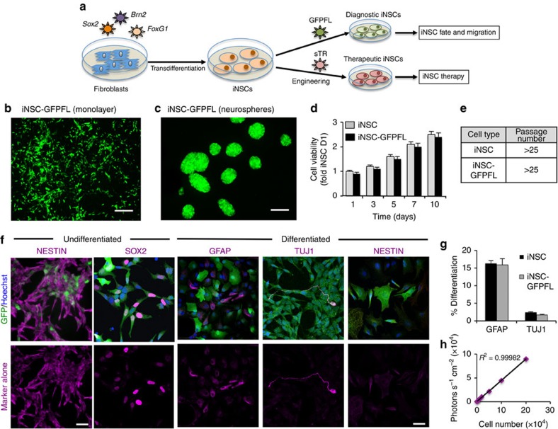
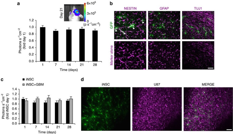
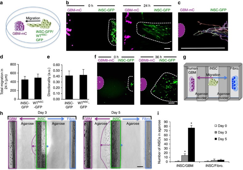
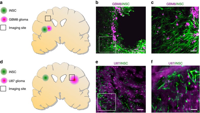
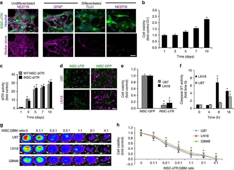
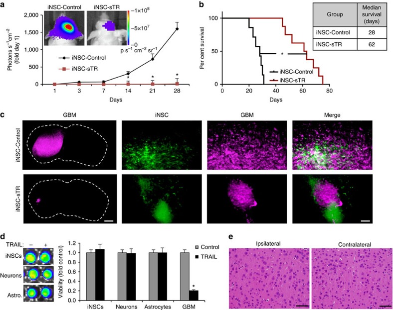
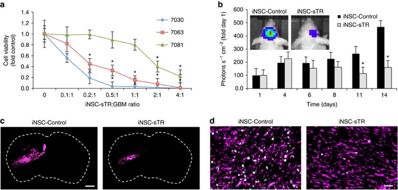
Transdifferentiation (TD) is a recent advancement in somatic cell reprogramming. The direct conversion of TD eliminates the pluripotent intermediate state to create cells that are ideal for personalized cell therapy. Here we provide evidence that TD-derived induced neural stem cells (iNSCs) are an efficacious therapeutic strategy for brain cancer. We find that iNSCs genetically engineered with optical reporters and tumouricidal gene products retain the capacity to differentiate and induced apoptosis in co-cultured human glioblastoma cells. Time-lapse imaging shows that iNSCs are tumouritropic, homing rapidly to co-cultured glioblastoma cells and migrating extensively to distant tumour foci in the murine brain. Multimodality imaging reveals that iNSC delivery of the anticancer molecule TRAIL decreases the growth of established solid and diffuse patient-derived orthotopic glioblastoma xenografts 230- and 20-fold, respectively, while significantly prolonging the median mouse survival. These findings establish a strategy for creating autologous cell-based therapies to treat patients with aggressive forms of brain cancer.


Figures

Figure 1
Figure 1. Generation and characterization of diagnostic and therapeutic iNSCs.
(a) Schematic depiction of the strategy used to create therapeutic and diagnostic variants of iNSCs. (b,c) Representative fluorescent photomicrographs of iNSCs engineered to express GFPFL and grown as monolayers (b) or neurospheres (c). (d) Summary graph showing the growth of GFPFL-expressing iNSCs in comparison with unmodified iNSCs. (e) Summary table showing the maximum passage number of iNSCs expressing GFPFL and unmodified iNSCs. (f) Representative images of immunofluorescence that show the expression of the NSC markers nestin and Sox2 (staining shown in magenta) in iNSC-GFPFL (green). In addition, iNSC-GFPFLs were differentiated by mitogen removal and culturing for 12 days. The cells were stained to detect GFAP+ astrocytes, Tuj-1+ neurons and nestin (staining shown in magenta). Fluorescent images showing only the red (555 nm) secondary antibody channel are shown in the bottom row. (g) Quantification of GFAP+ or Tuj-1+ cells present after differentiation of iNSC-GFPFL or unmodified iNSCs. (h) Summary data showing the linear correlation between iNSC-GFPFL cell number and bioluminescence signal. iNSC-GFPFLs were plated at increasing cell numbers, combined with D-luciferin and were imaged in a luminometer (R2=0.99982). Scale bars in b,c, 100 and 20 μm in f. Data are mean±s.e.m. Data in d,e,g,h are from three to four independent experiments. In d, P>0.05 by two-way ANOVA.
Figure 2
Figure 2. In vivocharacterization of iNSCs transplanted in the mouse brain in the presence or absence of GBM.
iNSC-GFPFLs were implanted into the frontal lobe of mice in the presence or absence of U87 human GBM (n=6). Serial bioluminescence imaging was used to monitor the volumes of iNSC-GFPFL. Two weeks after implantation, a subset of mice was killed and their brains sectioned and analysed. (a) Summary graph depicting the volume of iNSC-GFPFL in the brain in the absence of GBM through 28 days. (b) Immunofluorescence analysis of iNSC-GFPFL (green) 14 days post implantation into the brain. Nestin+ iNSC-GFPFLs were detected (indicated by arrowheads), and iNSC-GFPFL differentiation was detected with GFAP or Tuj-1 staining shown in magenta. Representative fluorescent images showing only the red (555 nm) secondary antibody channel are shown in the bottom row. (c) Engraftment of iNSC-GFPFL cells implanted in the brain in the presence or absence of human GBM (n=10 per group) measured using bioluminescence imaging. (d) Representative fluorescence imaging of post-mortem tissue sections showing GFP+ iNSCs (green) are still present in mCherry+ GBMs (magenta) 28 days after implantation. Scale bars in b,d, 40 and 50 μm, respectively. Data are mean±s.e.m. In a, P>0.05 by repeated measures one-way ANOVA. In c, P>0.05 by repeated measures two-way ANOVA.
Figure 3
Figure 3. Engineered iNSCs home to GBM.
(a) Outline of the iNSC migration assay. iNSC-GFPFLs were seeded 500 μm apart from mCherry+ human GBM cells and placed in a fluorescence incubator microscope. Time-lapse fluorescent images were captured every 10 min for 36 h and used to construct movies that revealed the migration of iNSCs in real time. (b) Summary images showing the migration of iNSC-GFPFL (green) towards U87-mC-FL (magenta) at 0 and 24 h after plating. (c) Single-cell tracings depicting the path of multiple iNSC-GFPFL migrating towards GBM cells over 24 h. Asterisk and dotted line indicate the site of GBM seeding. (d,e) Summary graph showing the total distance that iNSCs and brain-derived WTNSC migrated towards GBM cells (d), and directionality of iNSC-GFPFL and WTNSC migration to human U87 GBMs (e) determined from the real-time motion analysis. (f) Representative images showing the migration of iNSC-GFPFL to co-cultured patient-derived GBM8-mC cells (indicated by the dotted lines) at 0 and 36 h after plating. Asterisk and dotted line indicate the site of GBM8 seeding. (gi) Outline of agarose migration assays used to determine the selectivity of iNSC migration (g). Three wells were created in six-well culture plates containing agarose. iNSCs were seeded in the middle well, and GBM or fibroblasts were seeded in wells on either side. Real-time imaging (h) and summary data (i) show the number of iNSCs that migrated towards the wells containing GBM cells or the fibroblasts. Scale bars in b,f,h, 100 μm. Scale bar in c, 500 μm. Data are mean±s.e.m. Data in d,e,i are from three independent experiments. In d,e, P>0.05 by Student's t-test. In i, *P<0.05 by one-way ANOVA with Tukey's multiple comparisons test.
Figure 4
Figure 4. In vivo migration of iNSCs to GBMs.
(ac) To assess iNSC tracking of invading GBM cells, GBM8-mCs were implanted into the brains of mice (n=7). Three days later, iNSC-GFPFLs were implanted into the tumour. Twenty-one days later, fluorescent images of cryosections were captured 1.5 mm away (indicated by the square, a). (b,c) Representative fluorescent images showing the colocalization of iNSC-GFPFL (green) with invading GBM8-mC (magenta; b), and elongated iNSC morphology (c). (df) To assess the migration of iNSCs to distant established GBMs, mCherry+ U87 were implanted into the parenchyma of mice (n=7). iNSC-GFPFLs were implanted into the contralateral hemisphere 5 days later. Immunofluorescent analysis of post-mortem tissue sections 21 days post implant was used to determine the presence of GFP+ iNSCs (green) at the mCherry+ GBMs (magenta; square indicates site of imaging). Scale bars in b,e, 100 and 30 μm in c,f. Data are mean±s.e.m.
Figure 5
Figure 5. The antitumour efficacy of iNSC-sTR treatment of GBM cell lines in vitro.
(a) Representative images showing the expression of nestin, GFAP or Tuj-1 in undifferentiated and differentiated iNSC-sTR cells with IHC staining (iNSC-sTR=green; staining=magenta). Magenta images alone show only the fluorescent channel corresponding to nestin, GFAP and Tuj-1 staining. (b) Summary graph showing the viability of iNSC-sTR over 10 days determined using bioluminescence assays. (c) Summary data showing the levels of diTR released by iNSC-diTR or WTNSC-diTR cells determined with luciferase imaging on media samples. At each time point, equal volumes of media were collected from WTNSC-diTR or iNSC-diTR, combined with coelenterazine, and bioluminescence imaging was performed to determine the levels of secreted diTR. (d,e) Representative images (d) and summary graph (e) showing the cell viability of U87 and LN18 human GBM cells co-cultured with iNSC-sTR or iNSC-GFP assessed using luciferase-based assay. (f) Summary graphs showing caspase-3/7 activity in U87 and LN18 GBM cells treated with sTR or control conditioned media determined using a bioluminescence assays that incorporates a proluminescent caspase-3/7 substrate. (g,h) Representative bioluminescence imaging (g) and summary data (h) showing the viability of U87, LN18 and GBM8 tumour cells cultured with increasing numbers of iNSC-sTR. Scale bar in a, 20 and 50 μm in d. Data are mean±s.e.m. Data in b,c,e,f,h are from three to four independent experiments. In b, P>0.05 by one-way ANOVA. In c, P>0.05 by repeated measures two-way ANOVA. In e, *P<0.05 by Student's t-test. In f,h, *P<0.05 by repeated measures two-way ANOVA.
Figure 6
Figure 6. Antitumour efficacy of iNSC-sTR treatment of orthotopic human GBM xenografts.
Human U87 GBM cells expressing mCherry and firefly luciferase were implanted into the parenchyma of mice with iNSC-sTR or control iNSC-GFP. (a) Serial bioluminescence imaging showing the growth of the human GBM xenografts treated with iNSC-sTR or control iNSC-GFP through 28 days (n=12 per group). (b) Kaplan–Meier curves showing the survival of U87-bearing animals treated with iNSC-sTR or control iNSC (n=12 per group). (c) Representative fluorescent micrographs of brain sections from control- or iNSC-sTR-treated mice bearing mC-FL-expressing U87 tumours. Magenta=U87; Green=iNSC-sTR or iNSC-GFP. (d) Images and summary data showing the viability of iNSCs, neurons, astrocytes and U87 GBM cells cultured with conditioned media from iNSC-sTR or iNSC-GFP. (e) Representative histological images of the brain sections from mice injected intracranially with iNSC-sTR (n=5). Images were captured 0.5 mm from the implantation site (ipsilateral) and the same distance from the midline in the opposite hemisphere (contralateral). Scale bar in c, 1,000 and 30 μm in magnification. Scale bar e, 30 μm. Data are mean±s.e.m. Data in a,b,d are from three independent experiments. Data in a,b are n=12 per group. In a, *P<0.05 by repeated measures two-way ANOVA. In b, *P<0.01 by exact log-rank test. In d, *P<0.01 by Student's t-test.
Figure 7
Figure 7. Efficacy of iNSC-sTR therapy for highly invasive patient-derived GBM8 xenografts.
(a) Representative images of patient-derived GBM8 cancer cells expressing mC-FL (magenta) co-cultured with iNSC-sTR (green). (b) Summary data showing the cell viability of GBM8 cells after 24 h of co-culture with iNSC-sTR or control iNSCs assessed by luciferase assay. (c) mC-FL-expressing GBM8 glioma cells were incubated with conditioned media from iNSC-sTR or control cells, and caspase-3/7 activity was determined 18 h later. (dg) Highly invasive patient-derived GBM8 cells expressing mC-FL were implanted into the parenchyma of mice. Three days later, iNSC-sTRs or iNSC-GFPs were implanted into tumour. (d,e) Serial bioluminescence images tumour growth in iNSC-sTR- or iNSC-GFP-treated animals (n=12 per group). (f) Representative fluorescent micrographs of the brain sections from control- or iNSC-sTR-treated mice bearing mC-FL-expressing GBM8 tumours (magenta). (g) Kaplan–meier curves revealing the survival in animals treated with iNSC-sTR or iNSC-GFP (n=12 per group). Scale bar in a, 50 and 1,000 μm in f. Data are mean±s.e.m. Data in be are from three independent experiments. In b,c, *P<0.05 by Student's t-test. Data in d,g are n=12 per group. In b,c, *P<0.05 by paired t-test. In d, *P<0.05 by repeated measures two-way ANOVA. In g, *P<0.01 by exact log-rank test.
Figure 8
Figure 8. iNSC-sTR treatment of patient-derived GBMs.
(a) Summary data of the patient-derived cancer cells lines 7030, 7081, 7063, GBM6 treated with increasing volumes of conditioned media from iNSC-sTR or control iNSC-GFP cells. Cell viability was assessed 24 h after treatment. (b) Representative bioluminescent images and summary data showing the progression of 7063 xenografts treated with iNSC-sTR or iNSC-GFP (n=12 per group). (c) Representative fluorescent micrographs of the brain sections from control- or iNSC-sTR-treated mice bearing 7063 GBM xenografts (magenta). (d) Ki-67 (green) staining of post-mortem tissue sections 7063 (magenta) tumours treated with iNSC-control or iNSC-sTR. Colocalization of the signal is shown in white. Scale bar in c, 1,000 and 100 μm in d. Data in a are from three independent experiments. Data in b are n=12 per group. In a,b, *P<0.05 by repeated measures two-way ANOVA.

Comment in

Similar articles

Cited by

References

    1. Nakagawa M. et al. Generation of induced pluripotent stem cells without Myc from mouse and human fibroblasts. Nat. Biotechnol. 26, 101–106 (2008). - PubMed
    1. Okita K., Ichisaka T. & Yamanaka S. Generation of germline-competent induced pluripotent stem cells. Nature 448, 313–317 (2007). - PubMed
    1. Robinton D. A. & Daley G. Q. The promise of induced pluripotent stem cells in research and therapy. Nature 481, 295–305 (2012). - PMC - PubMed
    1. Lujan E. & Wernig M. The many roads to Rome: induction of neural precursor cells from fibroblasts. Curr. Opin. Genet. Dev. 22, 517–522 (2012). - PMC - PubMed
    1. Kim J. et al. Direct reprogramming of mouse fibroblasts to neural progenitors. Proc. Natl Acad. Sci. USA 108, 7838–7843 (2011). - PMC - PubMed

Publication types