

Prevalence of benign focal liver lesions: ultrasound investigation of 45,319 hospital patients
- PMID: 26830608
- PMCID: PMC4735268
- DOI: 10.1007/s00261-015-0605-7
Prevalence of benign focal liver lesions: ultrasound investigation of 45,319 hospital patients
Abstract
Purpose: The aim of the study was to determine the sonographic prevalence of benign focal liver lesions on the basis of a population of hospital patients.
Methods: The ultrasound results in a population of (n = 45,319) hospital patients over a period of 10 years were examined retrospectively and evaluated for the diagnosis of benign focal liver lesions [hepatic cysts, hepatic hemangioma, focal nodular hyperplasia (FNH), hepatic adenoma, and focal fatty sparing]. Results that were incomplete or ambiguous were excluded from this study.
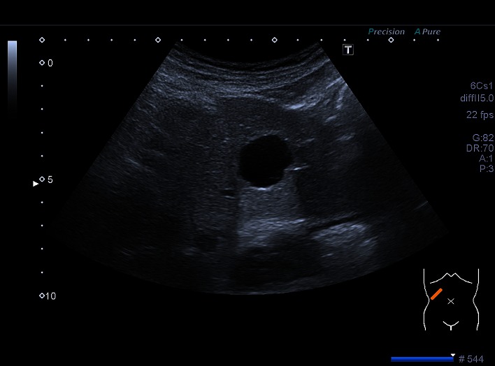
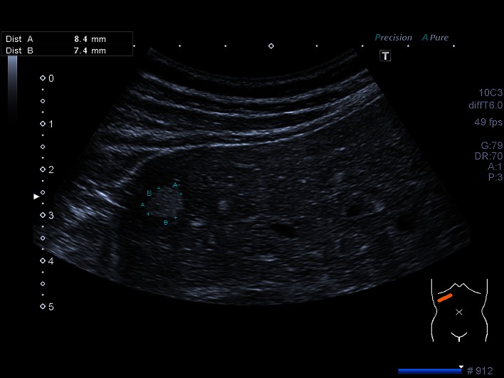
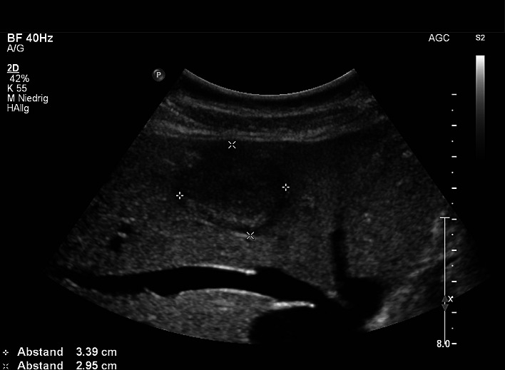
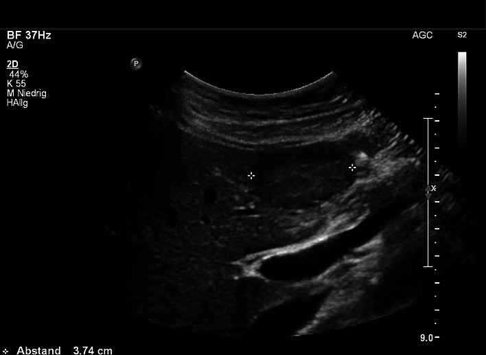
Results: At least one of the lesions to be investigated was diagnosed in 15.1% (n = 6839) of the patients of the total population. The most commonly recorded lesion, with a total prevalence of 6.3% (n = 2839), was focal fatty sparing, followed by hepatic cysts with 5.8% (n = 2631). The prevalence of hepatic hemangioma was 3.3% (n = 1640), while that of FNH was 0.2% (n = 81) and that of hepatic adenoma was 0.04% (n = 19). An association between the occurrence of benign focal liver lesions and age was observed.
Conclusions: The calculated prevalence of benign focal liver lesions shows that on the fortuitous discovery of space-occupying lesions of the liver, first consideration should be given to focal fatty sparing, simple hepatic cysts and hemangiomas. The finding of a FNH or an adenoma is rarely a random discovery.
Keywords: Focal fatty sparing; Focal liver lesions; Focal nodular hyperplasia; Hepatic adenoma; Hepatic cysts; Hepatic hemangioma.
Figures

References
MeSH terms
LinkOut - more resources
Full Text Sources
Other Literature Sources
Medical
