. 2016 Feb 1:6:20201.
doi: 10.1038/srep20201.

Glutamatergic axon-derived BDNF controls GABAergic synaptic differentiation in the cerebellum

Affiliations

Glutamatergic axon-derived BDNF controls GABAergic synaptic differentiation in the cerebellum

Albert I Chen et al. Sci Rep. .

Abstract

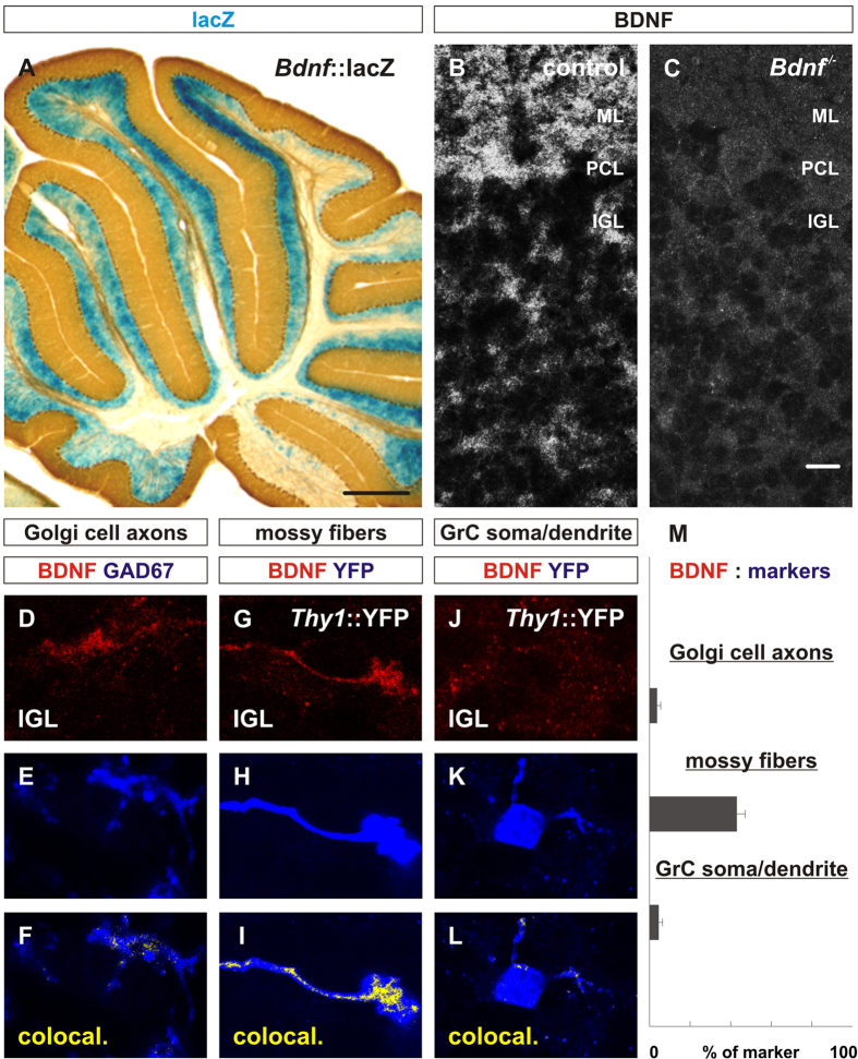
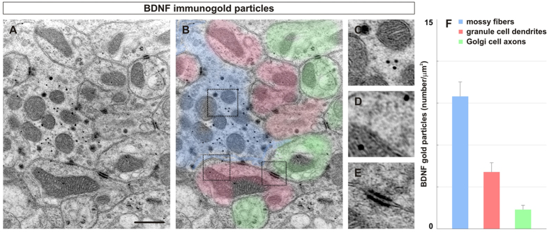
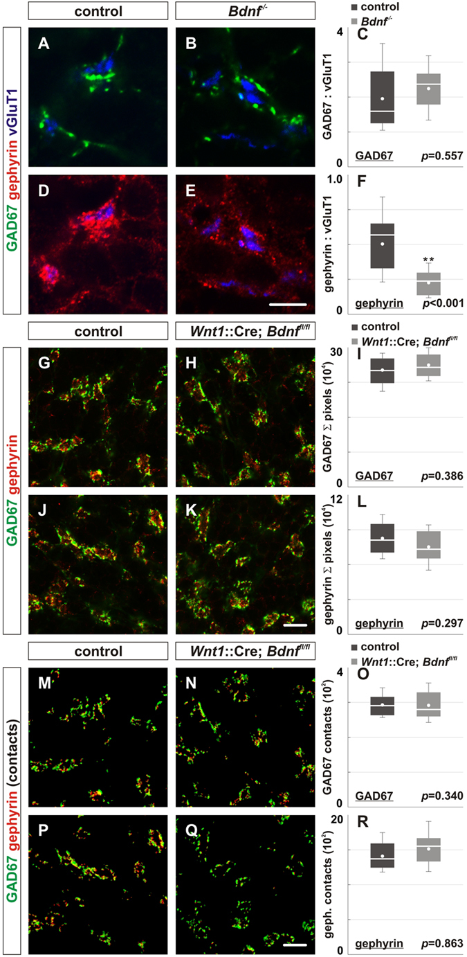
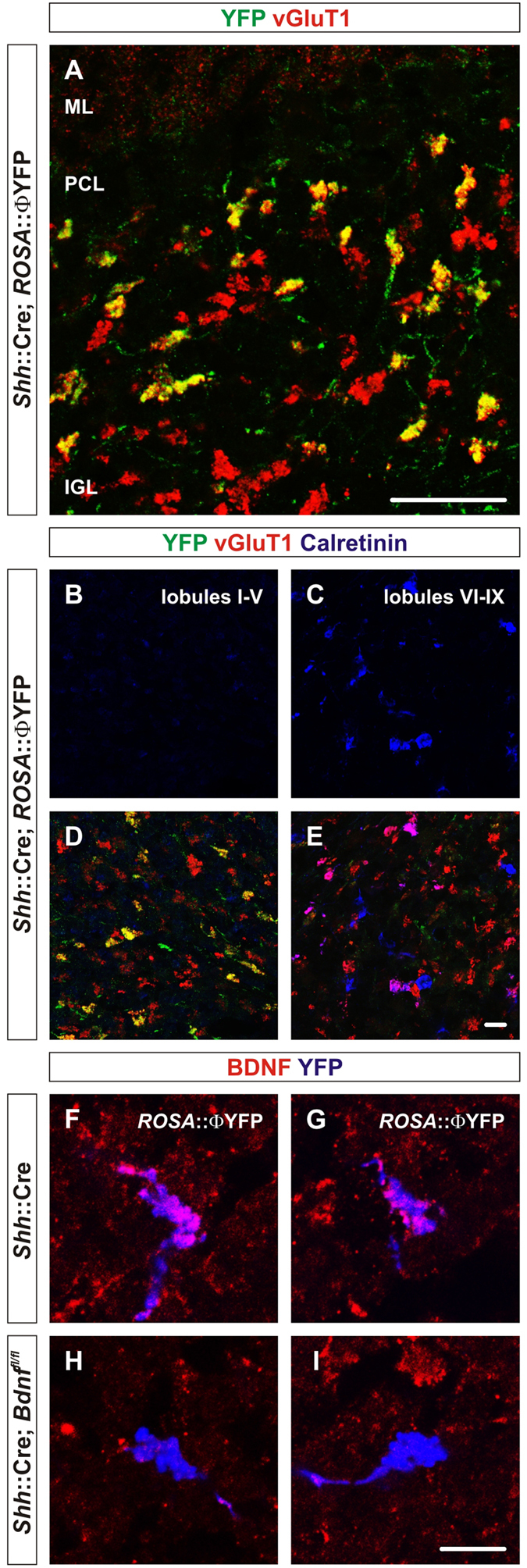
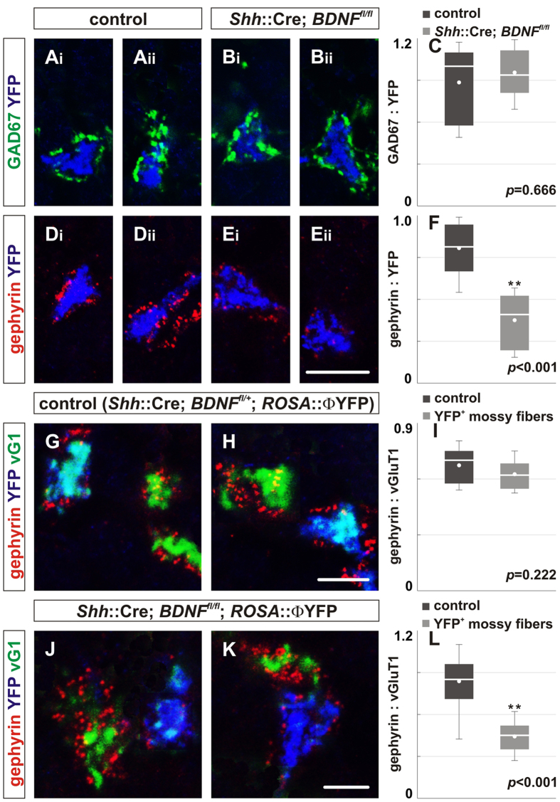
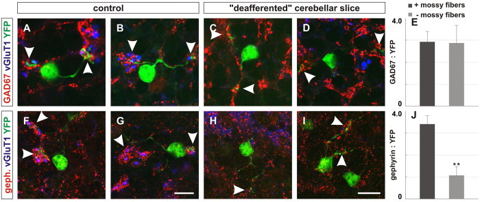
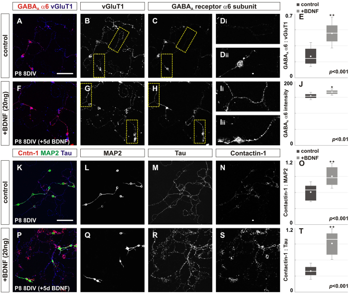
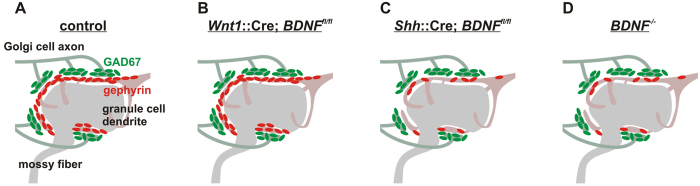
To study mechanisms that regulate the construction of inhibitory circuits, we examined the role of brain-derived neurotrophic factor (BDNF) in the assembly of GABAergic inhibitory synapses in the mouse cerebellar cortex. We show that within the cerebellum, BDNF-expressing cells are restricted to the internal granular layer (IGL), but that the BDNF protein is present within mossy fibers which originate from cells located outside of the cerebellum. In contrast to deletion of TrkB, the cognate receptor for BDNF, deletion of Bdnf from cerebellar cell bodies alone did not perturb the localization of pre- or postsynaptic constituents at the GABAergic synapses formed by Golgi cell axons on granule cell dendrites within the IGL. Instead, we found that BDNF derived from excitatory mossy fiber endings controls their differentiation. Our findings thus indicate that cerebellar BDNF is derived primarily from excitatory neurons--precerebellar nuclei/spinal cord neurons that give rise to mossy fibers--and promotes GABAergic synapse formation as a result of release from axons. Thus, within the cerebellum the preferential localization of BDNF to axons enhances the specificity through which BDNF promotes GABAergic synaptic differentiation.


Figures

Figure 1
Figure 1. Bdnf is expressed by cells in the internal granular layer and the protein is localized throughout the cerebellar cortex.
(A) The expression of BDNF is assessed by β-galactosidase reactivity in sagittal cerebellar sections obtained from three P30 BdnflacZ mice. LacZ expression is only detected in the internal granular layer (IGL) in sagittal cerebellar sections from BdnflacZ mice. (B,C) Using a monoclonal mouse antibody (Mab#9) against mature BDNF, we examined the localization of BDNF protein in three P14 Bdnf−/− mice compared to three control mice. (B) After antigen retrieval with sodium citrate, the BDNF protein is observed to be localized in the molecular layer, Purkinje cell layer and internal granular layer. (C) BDNF is lost in all layers of the cerebellar cortex in mice lacking Bdnf. (D–F) BDNF (red, E) is compared with GAD67 (blue, F) which labels Golgi cell terminals and does not appear to colocalize with Golgi cell axons (yellow, F). (G–I) When BDNF (red, G) is compared with vGluT1+ mossy fiber endings (blue, H), BDNF is extensively colocalized with mossy fiber endings (yellow, I). (J–L) BDNF (red, J) is compared with YFP (blue, K) which labels a subset of granule cells in Thy1::YFP mice. BDNF does not appear to colocalize with granule cell soma or dendrites (yellow, L). (M) Quantification of the area of BDNF on cell-type-specific markers indicate 4.33 ± 0.75% of GAD67+ Golgi cell axons contain BDNF, 43.08 ± 3.9% of vGluT1+ mossy fibers contain BDNF, 4.58 ± 0.84% of YFP+ granule cell soma/dendrites contain BDNF. Cerebella from four P21 wildtype mice and four P21 Thy1::YFP mice were used for analysis in D-M. Scale bar = 500 μm (A), 20 μm (B-P). *From herein, all statistics were done using Mann-Whitney U Test and errors represented as standard error of the mean.
Figure 2
Figure 2. Immunogold particles of BDNF in the internal granular layer are primarily localized on mossy fiber endings.
(A–E) We analyzed individual glomeruli from 20 electron micrographs obtained from four P60 wildtype mice (magnification 11,500X, single 2 μm sections) after immunogold labeling with BDNF Mab#9 antibody. (A–B) Individual components of a typical glomerulus (A) are color coded (B, mossy fibers = blue, granule cell dendrites = red, Golgi cell endings = green). (C–E) Regions correspond to dotted boxes. Most aggregates of gold grains are found in mossy fiber terminals (blue, B, C) while some gold grains are found in granule cell dendrites (red, B, D). Little gold clusters were found on Golgi cell axons (green, B, E). (F) Quantification of the number of gold particles per μm2 indicates 2.3X more gold particles were found in mossy fibers than in granule cell dendrites and 7.1X more gold particles than Golgi cell axons (MF = 9.41 ± 1.1/μm2 , granule cell = 4.08 ± 0.73/μm2, Golgi cell axons = 1.32 ± 0.31/μm2). Scale bar = 2 μm.
Figure 3
Figure 3. Consequences of complete versus specific deletion of Bdnf on the expression of GABAergic synaptic proteins and the number of contacts between these proteins in the internal granular layer.
(A–F) We analyzed the localization of pre- and postsynaptic GABAergic synaptic proteins in P14 conventional Bdnf−/− mice. (A,B) The expression of GAD67 (green) surrounding vGluT1+ mossy fibers (blue) is similar in Bdnf−/− mice (B) compared to control (A). (D,E) However, the expression of gephyrin (red) is significantly reduced in Bdnf−/− mice (E) compared to control (D). (C,F) Quantification of the area ratios of GAD67:vGluT1 and gephyrin:vGluT1 expression in Bdnf−/− mice (GAD67: 2.88 ± 0.79; gephyrin: 1.07 ± 0.37) compared to control (GAD67: 2.95 ± 0.48, p = 0.557; gephyrin: 3.4 ± 0.39, p < 0.001). (G–L) Deletion of Bdnf in the cerebellum did not influence GAD67 (green) or gephyrin (red) localization in the IGL of P21 Wnt1::Cre; Bdnffl/fl mice (G,J) compared to control (A,D). (I,L) Quantification of the area of the coverage of GAD67 (I) and gephyrin expression (L) in control (GAD67: 25.1 ± 0.9 × 104; gephyrin: 8.2 ± 0.5 × 104) and Wnt1::Cre; Bdnffl/fl mice (GAD67: 26.2 ± 0.8, p = 0.386; gephyrin: 7.4 ± 0.4 × 104, p = 0.297). (M–R) Similarly, loss of BDNF in the cerebellum did not influence the number of gephyrin contacts found on GAD67 puncta in the IGL of P21 Wnt1::Cre; Bdnffl/fl mice (N,Q) or the number of GAD67 contacts found on gephyrin puncta compared to control (M,P). (O,R) Quantification of the number of GAD67 contacts (# of gephyrin puncta on one GAD67 puncta) and the number of gephyrin contacts (# of GAD67 puncta on one gephyrin puncta) in control (GAD67 contacts: 2.79 ± 0.1 × 102; gephyrin contacts: 14.1 ± 0.7 × 102) and Wnt1::Cre; Bdnffl/fl mice (GAD67 contacts: 2.77 ± 0.02 × 102, p = 0.863; gephyrin contacts: 15.1 ± 0.7 × 102, p = 0.340). At least four mice were analyzed for each genotype described. Scale bar = 10 μm. *From herein, box plots contain lower and upper limits (“whiskers”), 1st and 3rd quartile (edges of the box), median (white bar), mean (white dot), and outliers (black dots) of the dataset.
Figure 4
Figure 4. Shh::Cre mice recombine in a subset of mossy fibers derived from precerebellar nuclei and/or spinal cord neurons.
(A) At P21, Shh::Cre mice selectively promote recombination in a subset of neurons that give rise to mossy fibers as assessed by YFP expression using the ROSA::ΦYFP reporter allele. Φ indicates a loxP-STOP-loxP cassette. Colocalization of vGluT1 expression with YFP+ mossy fibers (green) in P60 Shh::Cre; ROSA:: ΦYFP mice indicates that recombination occurs in a subset of mossy fibers (yellow). (B,C) The expression of Calretinin (blue) is mostly absent in lobule I-lobule V (B) and primarily found in lobule VI-lobule X (C). (D,E) The expression of YFP (green), which colocalizes with vGluT1, is found mostly in lobule I-lobule V (D), but not in lobule VI-lobule IX (E) in P60 Shh::Cre; ROSA::ΦYFP mice. Scale bar = 25 μm (A), 10 μm (B–E). (F–I) Shh::Cre mice effectively mediate deletion of BDNF in Shh::Cre; ROSA::ΦYFP; Bdnffl/fl mice. BDNF (red) is found in mossy fibers (blue) of Shh::Cre; ROSA::ΦYFP mice (F,G), but is absent in mossy fibers (blue) of Shh::Cre; ROSA::ΦYFP; Bdnffl/fl mice (H,I). Three mice of each genotype were analyzed. Scale bar = 50 μm (A), 10 μm (B–I). Φ indicates a loxP-STOP-loxP cassette.
Figure 5
Figure 5. Postsynaptic differentiation at GABAergic synapses depends on mossy fiber-derived BDNF signaling.
To explore the possibility that BDNF comes from an extracerebellar source, we deleted BDNF from a subset of mossy fibers and analyzed GABAergic synaptic proteins surrounding mossy fiber endings in the IGL. (A–F) YFP expression marks mossy fiber terminals from recombined neurons. Loss of BDNF in YFP+ mossy fiber endings did not influence GAD67 (green) localization in the IGL of P21 Shh::Cre; Bdnffl/fl; ROSA::ΦYFP mice (Bi, Bii) compared to control (Ai, Aii). However, loss of BDNF in YFP+ mossy fiber endings resulted in reduced gephyrin (red) localization in the IGL of P21 Shh::Cre; Bdnffl/fl; ROSA::YFP mice (Ei, Eii) compared to control (Di, Dii). (C,F) Quantification of the area ratio of GAD67:YFP (C) and the area ratio of gephyrin:YFP expression (F) in control (GAD67: 0.89 ± 0.09; gephyrin: 0.77 ± 0.05) and Shh::Cre; Bdnffl/fl; ROSA::ΦYFP mice (GAD67: 0.97 ± 0.06, p = 0.666; gephyrin: 0.36 ± 0.05, p < 0.001). (G–H) The localization of gephyrin (red) surrounding vGluT1 between YFP+ mossy fibers (teal) and unrecombined YFP- mossy fibers (green) is comparable in control mice. (I) Quantification of the area ratio of gephyrin:vGluT1 expression in P21 Shh::Cre; Bdnffll+; ROSA::ΦYFP mice (YFP- mossy fiber = 0.67 ± 0.01; YFP+ mossy fiber = 0.62 ± 0.01, p = 0.222). (J,K) However, the localization of gephyrin surrounding vGluT1 is reduced in recombined YFP+ mossy fibers (blue/teal) compared to unrecombined YFP- mossy fibers (green) in mice lacking BDNF in a subset of mossy fibers. (L) Quantification of the area ratio of gephyrin:vGluT1 expression in P21 Shh::Cre; Bdnffllfl; ROSA::ΦYFP mice (YFP- mossy fiber = 0.84 ± 0.02; YFP+ mossy fiber = 0.44 ± 0.01, p < 0.001). More than four mice were analyzed for each genotype. Scale bar = 10 μm.
Figure 6
Figure 6. Loss of mossy fiber endings in cultured cerebellar slices perturbs postsynaptic differentiation.
To assess whether the presence of mossy fibers or factors derived from mossy fibers regulate GABAergic synaptic differentiation, we generated cerebellar slices from P14 mice and cultured the slices in vitro for one week. (A–D,F–I) Cerebellar slices cultured in vitro for one week contain fewer vGluT1+ mossy fibers (blue) in the internal granular layer (C, D, H, I, white arrows) compared to age-matched control slices (A, B, F, G, white arrows). The localization of GAD67 (red) remains normal surrounding granule cell dendrites in “deafferented” cerebellar slices (C,D) compared to control (A,B). However, the localization of gephyrin (red) is significantly reduced in “deafferented” cerebellar slices (H, I) compared to control (F,G). (E,J) Quantification of the area ratios of GAD67:YFP and gephyrin:YFP expression in “deafferented” cerebellar slices (GAD67: 2.88 ± 0.79; gephyrin: 1.07 ± 0.37) compared to control slices (GAD67: 2.95 ± 0.48, p = 0.557; gephyrin: 3.4 ± 0.39, p < 0.001). At least four slices from three mice were analyzed for each condition. Scale bar = 20 μm.
Figure 7
Figure 7. Exogenous BDNF increases the expression of the α6 subunit of GABAA receptors and Contactin-1.
(A–J) Addition of recombinant BDNF in primary cerebellar granule cell culture promotes the expression of the α6 subunit of GABAA receptors. (A,F) Cerebellar granule cells are labeled by vGluT1 (blue) and the expression of GABAAR α6 subunit is shown in red. The expression of vGluT1 is not noticeably different after the addition of BDNF (F,G) compared to control (A,B). However, the expression of GABAAR α6 subunit is significantly increased after the treatment of BDNF for 5 days in culture (H, Ii, Iii) compared to control (C, Di, Dii). (E) Quantification of the area coverage of GABAAR α6 subunit over total vGluT1 in control (0.23 ± 0.13) and in culture after BDNF treatment (0.49 ± 0.17, p < 0.001). (J) Quantification of the intensity of GABAAR α6 subunit in control (155 ± 13) and in culture after BDNF treatment (168 ± 9.5, p < 0.01). (K–T) The expression of Contactin-1 on granule cell dendrites (MAP2, green, K, P) and axons (Tau, blue, K, P) was assessed following BDNF treatment. The expression of MAP2 is not noticeably different after the addition of BDNF (Q) compared to control (L), though the expression of Tau appears more punctate after the addition of BDNF (R) compared to control (M). However, the expression of Contactin-1 on the dendrites and axons of granule cells is significantly increased following BDNF treatment (S) compared to control (N). (O) Quantification of the area coverage of Contactin-1 over total MAP2 in control (0.61 ± 0.19) and in culture after BDNF treatment (0.91 ± 0.23, p < 0.001). (T) Quantification of the area coverage of Contactin-1 over total Tau in control (0.39 ± 0.15) and in culture after BDNF treatment (0.92 ± 0.28, p < 0.001). Scale bar = 50 μm.
Figure 8
Figure 8. Summary of the effects of BDNF deletion on GABAergic synaptic protein localization.
(A) The cerebellar glomerulus consists of mossy fiber terminals, granule cell dendrites and Golgi cell axons. GAD67 (green), a presynaptic GABAergic protein localized on Golgi cell axon endings, is adjacent to gephyrin (red), a postsynaptic GABAergic protein localized on granule cell dendrites. (B) Deletion of BDNF in the cerebellum did not result in any changes in the localization of GAD67 or gephyrin at the cerebellar glomerulus. (C,D) However, deletion of BDNF in mossy fibers (C) or conventional deletion of BDNF (D) resulted in reduced localization of gephyrin without any effects on the localization of GAD67.

Similar articles

Cited by

References

    1. Chao M. V., Rajagopal R. & Lee F. S. Neurotrophin signalling in health and disease. Clinical Sci 110, 167–173 (2006). - PubMed
    1. Bloodgood B. L. et al. The activity-dependent transcription factor NPAS4 regulates domain-specific inhibition. Nature 503, 121–125 (2013). - PMC - PubMed
    1. Je H. S. et al. ProBDNF and mature BDNF as punishment and reward signals for synapse elimination at mouse neuromuscular junctions. J Neurosci 33, 9957–9962 (2013). - PMC - PubMed
    1. Kaneko M., Xie Y., An J. J., Stryker M. P. & Xu B. Dendritic BDNF synthesis is required for late-phase spine maturation and recovery of cortical responses following sensory deprivation. J Neurosci 32, 4790–4802 (2012). - PMC - PubMed
    1. Nagappan G. et al. Control of extracellular cleavage of ProBDNF by high frequency neuronal activity. Proc Natl Acad Sci USA 106, 1267–1272 (2009). - PMC - PubMed

Publication types

MeSH terms