. 2016 Feb 2:6:20084.
doi: 10.1038/srep20084.

Additive enhancement of wound healing in diabetic mice by low level light and topical CoQ10

Affiliations

Additive enhancement of wound healing in diabetic mice by low level light and topical CoQ10

Zhigang Mao et al. Sci Rep. .

Abstract

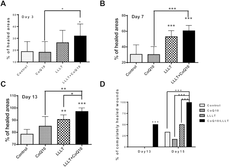
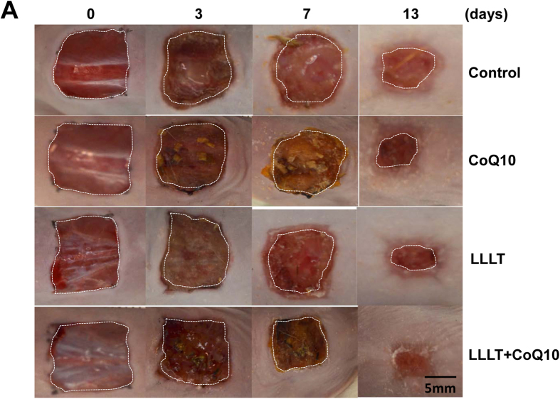
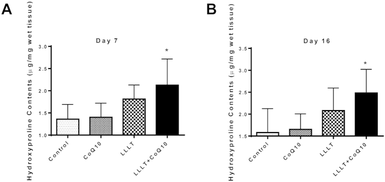
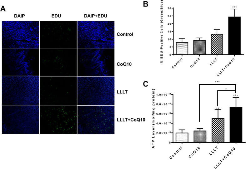
Diabetes, a highly prevalent disease that affects 9.3% of Americans, often leads to severe complications and slow wound healing. Preclinical studies have suggested that low level light therapy (LLLT) can accelerate wound healing in diabetic subjects, but significant improvements must be made to overcome the absence of persuasive evidence for its clinical use. We demonstrate here that LLLT can be combined with topical Coenzyme Q10 (CoQ10) to heal wounds in diabetic mice significantly faster than LLLT alone, CoQ10 alone, or controls. LLLT followed by topical CoQ10 enhanced wound healing by 68~103% in diabetic mice in the first week and more than 24% in the second week compared with untreated controls. All wounds were fully healed in two weeks following the dual treatment, in contrast to only 50% wounds or a fewer being fully healed for single or sham treatment. The accelerated healing was corroborated by at least 50% higher hydroxyproline levels, and tripling cell proliferation rates in LLLT and CoQ10 treated wounds over controls. The beneficial effects on wound healing were probably attributed to additive enhancement of ATP production by LLLT and CoQ10 treatment. The combination of LLLT and topical CoQ10 is safe and convenient, and merits further clinical study.


Figures

Figure 1
Figure 1. Additive effects of LLLT and CoQ10 on wound healing in diabetic mice.
Cutaneous full-thickness wounds were generated in diabetic mice and treated 24 hr later with sham light plus topical vehicle of CoQ10 (control), sham light + topical CoQ10 (CoQ10), LLLT+vehicle (LLLT), or LLLT followed by topical CoQ10 (LLLT+CoQ10). Wound areas were measured with Image J of photos taken at indicated days post-injury (A–C). The level of wound healing was defined as percentages of re-epithelialized areas over original wound areas as detailed in Materials and Methods. (D) Percentages of wounds with complete healing in each group. Complete wound healing as defined by full epithelialization was tracked in 13 and 15 days. Note: all skin wounds were completely healed in day 15 in LLLT+CoQ10 group only. *p < 0.05, **p < 0.01 and ***p < 0.001 between LLLT+CoQ10 group and all other three groups or between indicated groups. n = 12 in each group for data collected before or on day 7 or n = 6 for data collected after day 7.
Figure 2
Figure 2. Representative photos of each treatment.
Wounds were generated and photos were taken as in Fig. 1. Each row depicts the same wound as it progresses for 13 days. Shown are representative photos from indicated treatments as above. Bar, 5 mm.
Figure 3
Figure 3. Increased hydroxyproline contents in the wound tissue after treatment with LLLT plus CoQ10.
Hydroxyproline contents were measured as μg/mg wet tissue in 7 (A) or 16 (B) days post-injury. *p < 0.05 between LLLT+CoQ10 group and all other three groups. n = 6 in each group.
Figure 4
Figure 4. Active cell proliferation in wound tissues following the dual treatment.
Cell proliferation was evaluated 7 days after wounds by the EdU assay. (A) Confocal microscopy of EdU-stained tissue sections. Green fluorescence represents EdU-positive cells and DAPI stained all cells. Representative results of 36 sections in each group are shown. Magnification, ×20. (B) EdU-fluorescence intensity. Percentages of EDU-positive cells over a total number of cells are determined by counting individual cells in 100 microscopic fields selected randomly. ***p < 0.001 between LLLT+CoQ10 group and all other three groups (n = 100). (C) ATP levels were measured in duplicate 1 hr after the first indicated treatments. *p < 0.05, **p < 0.01 and ***p < 0.001 between LLLT+CoQ10 group and all other three groups or between indicated groups. n = 8 in each group.

Similar articles

Cited by

References

    1. Al-Watban F., Zhang X. & Andres L. Low-level laser therapy enhances wound healing in diabetic rats: a comparison of different lasers. Photomed. Laser Surg. 25, 72–77 (2007). - PubMed
    1. Beckmann H., Meyer-Hamme G. & Schröder S. Low level laser therapy for the treatment of diabetic foot ulcers: a critical survey. Evidence-based Complement. Altern. Med. 626127, 10.1155/2014/626127 (2014). - DOI - PMC - PubMed
    1. Singh N., Armstrong D. & Lipsky A. Preventing foot ulcers in patients with diabetes. JAMA 293, 217–228 (2005). - PubMed
    1. Schultz S. et al.. Wound bed preparation: A systematic approach to wound management. Wound Repair Regen. 11, Suppl 1, S1–28. (2003). - PubMed
    1. Richard J., Sotto A. & Lavigne J. New insights in diabetic foot infection. World J. Diabetes 2, 24–32 (2011). - PMC - PubMed

Publication types