. 2016 Feb 11;164(4):770-9.
doi: 10.1016/j.cell.2016.01.011. Epub 2016 Jan 28.

Precision Tumor Recognition by T Cells With Combinatorial Antigen-Sensing Circuits

Affiliations

Precision Tumor Recognition by T Cells With Combinatorial Antigen-Sensing Circuits

Kole T Roybal et al. Cell. .

Abstract

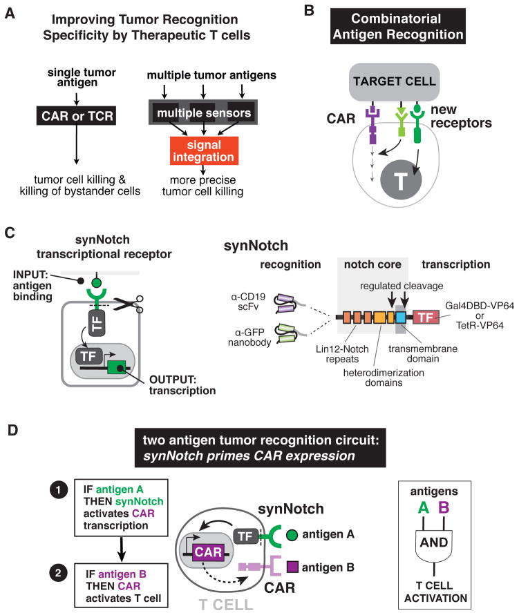
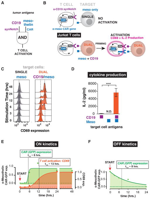
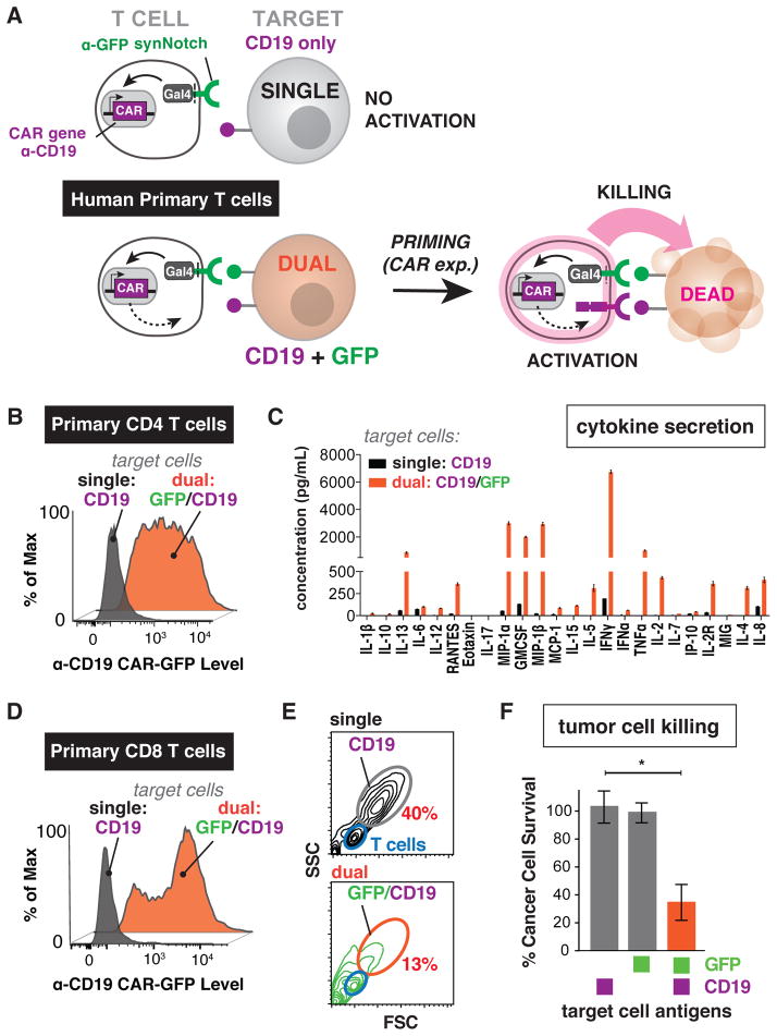
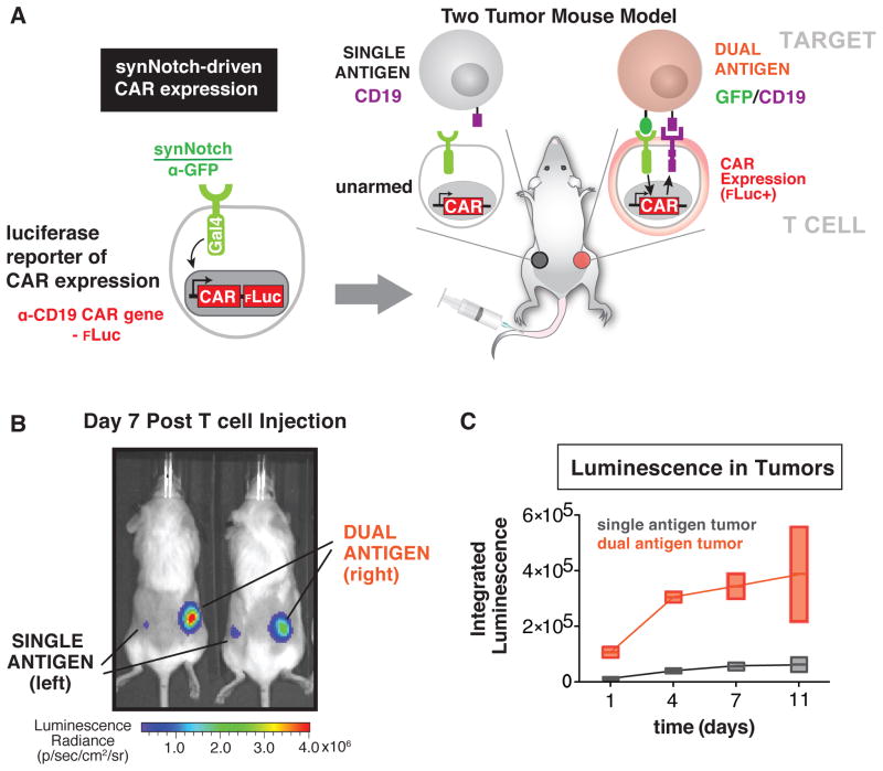
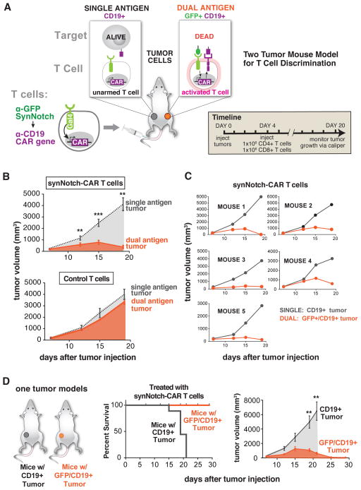
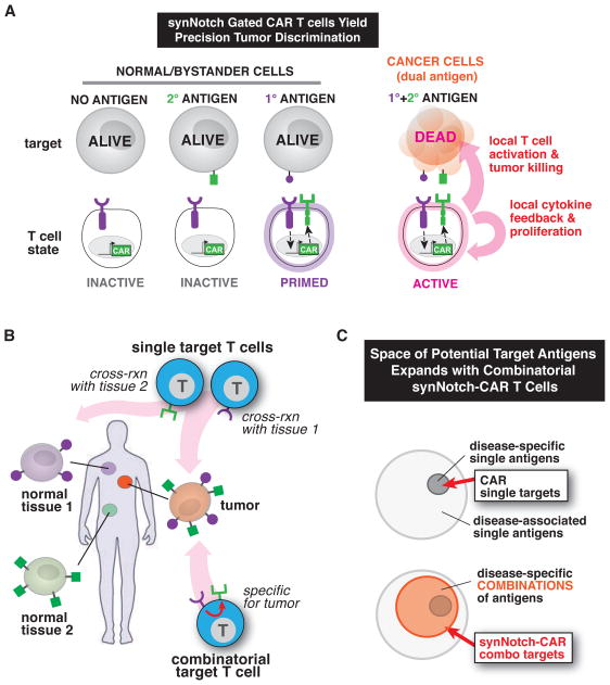
T cells can be re-directed to kill cancer cells using chimeric antigen receptors (CARs) or T cell receptors (TCRs). This approach, however, is constrained by the rarity of tumor-specific single antigens. Targeting antigens also found on bystander tissues can cause life-threatening adverse effects. A powerful way to enhance ON-target activity of therapeutic T cells is to engineer them to require combinatorial antigens. Here, we engineer a combinatorially activated T cell circuit in which a synthetic Notch receptor for one antigen induces the expression of a CAR for a second antigen. These dual-receptor AND-gate T cells are only armed and activated in the presence of dual antigen tumor cells. These T cells show precise therapeutic discrimination in vivo-sparing single antigen "bystander" tumors while efficiently clearing combinatorial antigen "disease" tumors. This type of precision dual-receptor circuit opens the door to immune recognition of a wider range of tumors. VIDEO ABSTRACT.


Figures

Figure 1
Figure 1. Design of Combinatorial Antigen Sensing Circuits in T cells Using Sequentially Regulated SynNotch and Chimeric Antigen Receptors
(A) CAR or tumor-specific TCR T cells generally target single antigens often causing OFF-target tissue damage. Improved therapeutic T cells will require multiple sensors that recognize combinations of both tumor antigens and tissue-specific antigens, allowing the cells to assess their environment and make more precise decisions on when to activate. Such therapeutic cells would be better equipped to distinguish target diseased tissue from normal tissue. (B) New types of receptors that sense combinations of antigens and regulate T cell signaling and transcription must be built to allow for sophisticated cellular decision-making and more precise therapeutic T cell responses. (C) SynNotch receptors are engineered with a custom extracellular ligand-binding domain (e.g. scFv or nanobody) directed towards an antigen of interest (e.g. CD19 or surface GFP). Upon ligand recognition by the synNotch receptor, an orthogonal transcription factor (e.g. TetR-VP64 or Gal4-VP64) is cleaved from the cytoplasmic tail that regulates a custom genetic circuit. (D) Design of a synNotch AND-gate circuit that requires T cells to sense two antigens to activate. This AND-gate signaling circuit works in two sequential steps: 1) A synNotch receptor allows the T cell to recognize the first antigen A, and 2) the T cell expresses a CAR directed towards a second tumor antigen B. If A and B are present, the T cells can activate and kill the target tumor.
Figure 2
Figure 2. SynNotch Regulated CAR Expression – Combinatorial Antigen Requirement for Jurkat T cell Activation
(A) Engineering a two-receptor AND-gate circuit: α-CD19 synNotch receptor induces α-mesothelin CAR expression. (B) Jurkat T cells were engineered with the α-CD19 synNotch tTa receptor and the corresponding response elements controlling α-mesothelin 4-1BBζ CAR expression. The Jurkat T cells must first recognize CD19 on the target tumor via their synNotch receptor to initiate CAR expression. After the T cell is primed to activate by CD19, the α-mesothelin CAR can then bind mesothelin and activate the Jurkat cell. Two canonical markers of T cell activation are CD69 upregulation and IL-2 production. The synNotch AND gate Jurkat T cells should only activate when exposed to target tumor cells expressing both CD19 and mesothelin. (C) Histograms of the activation marker CD69 in synNotch AND-gate Jurkat T cells co-cultured with single antigen (mesothelin only) or dual antigen (CD19/mesothelin) K562 tumor cells over a 48-hour timecourse. CD69 was only expressed when the T cells were exposed to dual antigen K562 cells (representative of 3 independent experiments). (D) IL-2 ELISA showing IL-2 production by synNotch AND-gate Jurkat cells only when exposed to dual antigen K562 cells (n=3, error bars are SEM, significance determined by Student’s t-test, **** = P ≤ 0.0001) (E) Timecourse of AND-gate T cell activation upon stimulation with dual antigen K562 cells. Expression of the GFP-tagged mesothelin CAR (green) occurs with a half-time of ~6 hours. Subsequently, activation of the T cell by CAR activation (monitored by CD69 expression) then occurs with a lag of several more hours (t1/2 ~13 hrs). FACS histograms for CAR expression are shown in Figure S1B. (F) Timecourse of AND-gate T cell inactivation upon removal of synNotch ligand. Jurkat T cells expressing the AND-gate circuit were stimulated for 24 hours by plate-bound α-Myc antibody (synNotch receptor has extracellular Myc-tag). START indicates time at which cells were removed from the ligand, and the decay of GFP tagged CAR expression was monitored (t1/2 ~8 hrs). FACS histograms for CAR expression are shown in Figure S1C.
Figure 3
Figure 3. SynNotch Regulated CAR Expression in Human Primary T cells – Combinatorial Antigen Control Over Therapeutic T cell Activation and Tumor Killing
(A) Human primary CD4+ and CD8+ T cells were engineered with the α-GFP nanobody synNotch Gal4VP64 receptor and the corresponding response elements controlling expression of the α-CD19 4-1BBζ CAR. These CD4+ or CD8+ synNotch AND gate T cells first must sense surface GFP via their synNotch receptor and only then do they express the α-CD19 CAR and are primed to activate. These AND-gate primary T cells should only activate and produce cytokine or kill target cells if they sense both GFP and CD19. (B) Primary CD4+ synNotch AND gate T cells described in panel A were co-cultured with CD19 only or surface GFP/CD19 K562 cells. Histograms of α-CD19 CAR GFP receptor expression level show that the CAR is only expressed when GFP is present on the surface of the target cell (representative of at least 3 independent experiments). (C) The supernatant from CD4+ synNotch AND gate T cells activated either by CD19 only or GFP/CD19 K562s was analyzed for the presence of 25 cytokines via Luminex. Cytokines were only produced when the T cells were exposed to GFP/CD19 T cells (error bars are SEM, n=3). (D) CD8+ synNotch AND gate primary T cells were engineered as described in panel A. As with the CD4+ T cells, the histograms of α-CD19 CAR GFP receptor expression level show that the CAR is only expressed when GFP is present on the surface of the target cell (representative of at least 3 independent experiments). (E) Forward and side scatter flow cytometry plots after 24 hour co-culture of CD8+ synNotch AND gate primary T cells with either CD19 only or GFP/CD19 tumors cells. The T cells fall within the blue gate and the target CD19 or the GFP/CD19 K562s are in the gray and orange gates, respectively. The synNotch AND gate T cells only killed the GFP/CD19 K562s, shown by the reduction of cells in the K562 gate (representative of 3 experiments). (F) Quantification of replicate CD8+ synNotch AND gate primary T cell cytotoxicity data shown in panel E. (n=3, error bars are SEM, significance determined by Student’s t-test * = P ≤ 0.05). Other examples of synNotch→CAR circuits in primary T cells are shown in Figure S2.
Figure 4
Figure 4. SynNotch Receptors Drive Tumor Localized CAR Expression In vivo
(A) Primary human CD4+ and CD8+ T cells were engineered with the α-GFP synNotch Gal4VP64 receptor and the corresponding response elements regulating α-CD19 4-1BBζ CAR IRES effluc expression and injected i.v. into NSG mice with a Daudi tumor (CD19 only) on the left flank and a surface GFP Daudi (GFP/CD19) tumor on the right flank. Luciferase expression was monitored over 11 days after i.v. injection of engineered T cells. (B) A representative image of luciferase expression in mice treated as described in panel A at day 7 post T cell injection. Luciferase expression was high in the GFP/CD19 tumor indicating localized CAR expression only in the dual antigen tumor (n=2 mice). (C) Quantification of integrated intensity of luciferase levels in the left flank Daudi tumor (CD19 only) and surface GFP Daudi tumor (GFP/CD19) in the right flank. Luciferase expression is enriched in the dual antigen tumor at all time points (error is SD n=2).
Figure 5
Figure 5. Selective Combinatorial Antigen Tumor Killing In vivo by SynNotch Gated CAR Expression
(A) Primary human CD4+ and CD8+ T cells were engineered with the α-GFP synNotch Gal4VP64 receptor and the corresponding response elements regulating α-CD19 4-1BBζ CAR expression and were injected i.v. into NSG mice with a CD19 K562 tumor on the left flank and a surface GFP/CD19 K562 tumor on the right flank. Tumor size was monitored over 16 days after i.v. injection of engineered T cells or untransduced T cell controls. (B) Graphs showing CD19 and GFP/CD19 tumor volumes for mice treated with synNotch AND gate T cells (top) and untransduced control T cells (bottom). synNotch AND gate T cells target the dual antigen tumor exclusively and the CD19 only tumor grew at the same rate as in mice treated with untransduced control T cells (n=5 mice, error bars are SEM, significance determine by Student’s t-test ** = P ≤ 0.01, *** = P ≤ 0.001). (C) Tumor volume measurement for individual mice treated with synNotch AND gate T cells. All mice showed selective killing of the dual antigen tumor. (D) Kaplan-Meier graphs showing synNotch AND gate T cells clear GFP/CD19 tumors with 100% of the mice surviving. Mice with CD19 only tumors are not cleared by synNotch AND gate T cells and have uncontrolled tumor growth. The corresponding tumor growth curves are given on the right of panel D (n=5 mice, error bars are SEM, significance determine by Student’s t-test ** = P ≤ 0.01).
Figure 6
Figure 6. SynNotch Receptors Control and Localize CAR T cell Response for Precision Immunotherapy
(A) Here we engineered T cells with synNotch receptors that sense tumor antigens and upregulate expression of a CAR to a second antigen. Thus, these synNotch AND gate T cells only activate in response to combinatorial antigen recognition in the tumor microenvironment, preventing OFF-target toxicity mediated by single antigen recognition. (B) SynNotch AND gate T cells, unlike therapeutic T cells that target single antigens, can reliably discriminate combinatorial antigen targets from single antigen bystander tissue. Combinatorial antigen sensing by synNotch-CAR T cells could aid in precisely targeting T cells to tumors preventing OFF-target toxicity. (C) synNotch receptors expand the targetable tumor antigen space. Tumor-specific antigens are rare compared to tumor-associated antigens (antigens that are expressed on normal tissue but are more highly expressed on tumors). Since CARs fully activate T cells resulting in the killing of target tissue, T cells engineered with a single CAR must be targeted to tumor-specific antigens in order to reduce fatal OFF-target toxicity (top venn diagram). SynNotch receptors can gate CAR expression and control where the T cells are armed. When targeting tumor specific antigen combinations, it may now be possible to use CAR receptors directed towards tumor-associated antigens. This should reduce OFF-target damage to tissues that express the CAR antigen in other parts of the body.

Comment in

Similar articles

Cited by

References

    1. Barrett DM, Singh N, Porter DL, Grupp SA, June CH. Chimeric antigen receptor therapy for cancer. Annu Rev Med. 2014a;65:333–347. - PMC - PubMed
    1. Barrett DM, Teachey DT, Grupp SA. Toxicity management for patients receiving novel T-cell engaging therapies. Curr Opin Pediatr. 2014b;26:43–49. - PMC - PubMed
    1. Brentjens RJ, Davila ML, Rivière I, Park J, Wang X, Cowell LG, Bartido S, Stefanski J, Taylor C, Olszewska M, et al. CD19-targeted T cells rapidly induce molecular remissions in adults with chemotherapy-refractory acute lymphoblastic leukemia. Sci Transl Med. 2013;5:177ra38–177ra38. - PMC - PubMed
    1. Chen L, Flies DB. Molecular mechanisms of T cell co-stimulation and co-inhibition. Nat Rev Immunol. 2013;13:227–242. - PMC - PubMed
    1. Chinnasamy D, Yu Z, Theoret MR, Zhao Y, Shrimali RK, Morgan RA, Feldman SA, Restifo NP, Rosenberg SA. Gene therapy using genetically modified lymphocytes targeting VEGFR-2 inhibits the growth of vascularized syngenic tumors in mice. J Clin Invest. 2010;120:3953–3968. - PMC - PubMed

Publication types