. 2016;36(3):261-70.
doi: 10.3109/10799893.2015.1091475. Epub 2015 Sep 29.

α1A-Adrenergic receptor prevents cardiac ischemic damage through PKCδ/GLUT1/4-mediated glucose uptake

Affiliations

α1A-Adrenergic receptor prevents cardiac ischemic damage through PKCδ/GLUT1/4-mediated glucose uptake

Ting Shi et al. J Recept Signal Transduct Res. 2016.

Abstract

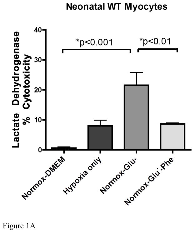
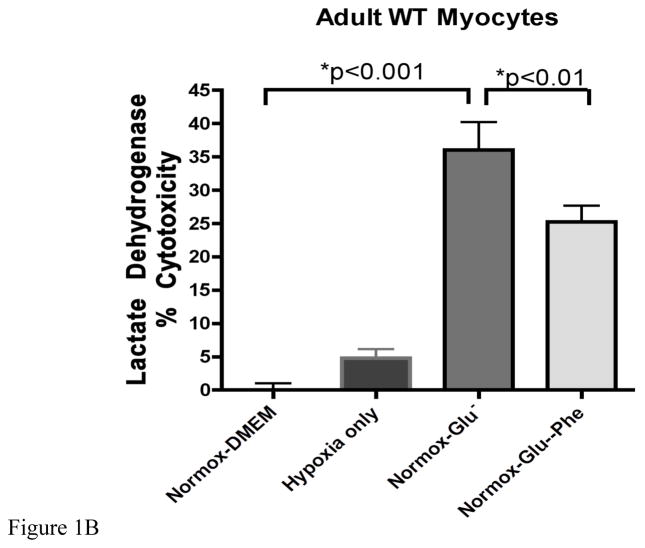
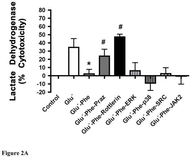
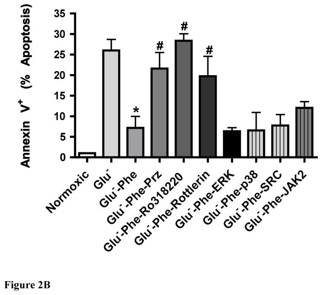
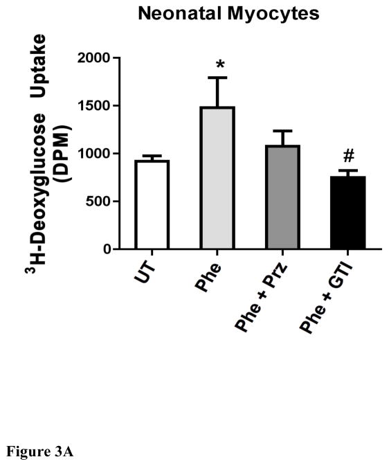
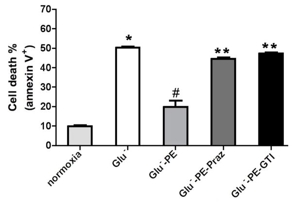
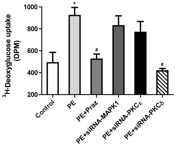
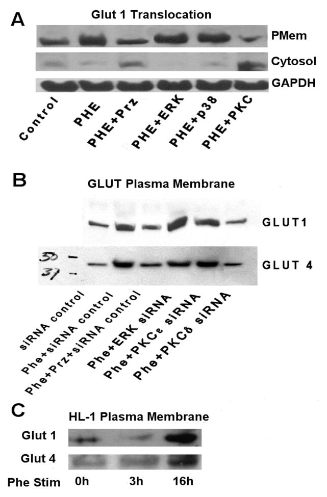
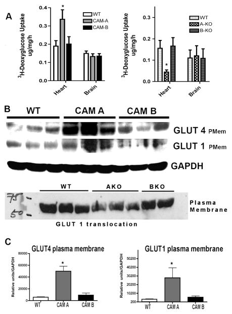
While α(1)-adrenergic receptors (ARs) have been previously shown to limit ischemic cardiac damage, the mechanisms remain unclear. Most previous studies utilized low oxygen conditions in addition to ischemic buffers with glucose deficiencies, but we discovered profound differences if the two conditions are separated. We assessed both mouse neonatal and adult myocytes and HL-1 cells in a series of assays assessing ischemic damage under hypoxic or low glucose conditions. We found that α(1)-AR stimulation protected against increased lactate dehydrogenase release or Annexin V(+) apoptosis under conditions that were due to low glucose concentration not to hypoxia. The α(1)-AR antagonist prazosin or nonselective protein kinase C (PKC) inhibitors blocked the protective effect. α(1)-AR stimulation increased (3)H-deoxyglucose uptake that was blocked with either an inhibitor to glucose transporter 1 or 4 (GLUT1 or GLUT4) or small interfering RNA (siRNA) against PKCδ. GLUT1/4 inhibition also blocked α(1)-AR-mediated protection from apoptosis. The PKC inhibitor rottlerin or siRNA against PKCδ blocked α(1)-AR stimulated GLUT1 or GLUT4 plasma membrane translocation. α(1)-AR stimulation increased plasma membrane concentration of either GLUT1 or GLUT4 in a time-dependent fashion. Transgenic mice overexpressing the α(1A)-AR but not α(1B)-AR mice displayed increased glucose uptake and increased GLUT1 and GLUT4 plasma membrane translocation in the adult heart while α(1A)-AR but not α(1B)-AR knockout mice displayed lowered glucose uptake and GLUT translocation. Our results suggest that α(1)-AR activation is anti-apoptotic and protective during cardiac ischemia due to glucose deprivation and not hypoxia by enhancing glucose uptake into the heart via PKCδ-mediated GLUT translocation that may be specific to the α(1A)-AR subtype.

Keywords: Adrenergic receptor; cardiac; glucose transporter.


Conflict of interest statement

Declaration of Interest: The authors report no conflicts of interest. This work was supported by The Heart Lung Blood Institute from The National Institutes of Health [RO1HL098279] and an American Heart Association Grant in Aid (Great Rivers Affiliate) to DMP.

Figures

Figure 1
Figure 1. α1-AR stimulation reduced lactate dehydrogenase release in wild-type neonatal myocytes undergoing glucose but not oxygen deprivation
A. Neonatal myocytes were subjected to normoxic (normox; atmospheric O2) or hypoxia (1% O2 for 24 hr) as described in materials and methods with either normal glucose (DMEM, 22.5mM) or low glucose concentrations (Glu, 1.375 mM) with or without α1-AR stimulation (Phe; 100μM). B. Adult myocytes were subjected to same conditions as above except incubation time was 5 hours. The amount of LDH released (% cytotoxicity) into the media was measured using the LDH Cytotoxicity Detection Kit (Clontech Laboratories, Inc., Mountain View, CA). *Statistically significant. N = 4–6 independent experiments were performed in triplicate.
Figure 2
Figure 2. α1-AR stimulation reduced lactate dehydrogenase release (A) or annexin V+ apoptosis (B) in WT neonatal myocytes undergoing glucose deprivation through a PKC-mediated pathway
Neonatal myocytes seeded onto 96-well plates were subjected to normoxic conditions (control) or with low glucose concentrations (Glu; 1.375mM) with or without α1-AR stimulation (Phe; 100μM). α1-AR stimulated cells were incubated with a series of inhibitors: prazosin, Praz (1μM); protein kinase C, PKC (1.5μM Ro-31-8220 or 1.25μM rottlerin), ERK (25μM PD98059), p38 (10μM SB203580), SRC (10μM PP2) and JAK2 (25μM AG490). *Statistically significant p≤0.05 from Glu−. #Statistically significant p≤0.05 from Glu-Phe. N = 5 independent experiments performed in triplicate.
Figure 3
Figure 3. α1-AR stimulation increased deoxyglucose uptake in either neonatal (A) or adult (B) myocytes
Cells were seeded at 60–80% confluence, incubated overnight, then switch to serum free medium for 2–4 hr. Cells were treated with or without (untreated, UT) phenylephrine (PE, 100 μM) for 16hr and either prazosin (Praz, 1 μM) or the glucose transporter inhibitor II (GTI, 1μM). [3H]-2-deoxyglucose (2DG) was added to each well (0.5 μCi/well) and incubated for 10 minutes. The cells were washed, lysed and transferred to a scintillation vial where 3H-2DG uptake was detected using Beckman scintillation counter. *Statistically significant p≤0.05 from untreated control. #Statistically significant p≤0.05 from phenylephrine stimulation. N = 5 independent experiments performed in triplicate.
Figure 4
Figure 4. α1-AR mediated glucose uptake protected against annexin V+ apoptosis in HL-1 cells
Cells (105) were subjected to normoxic conditions (control) or with low glucose concentrations (Glu; 1.375mM) with or without α1-AR stimulation (PE; 100μM). α1-AR stimulated cells were incubated with prazosin, Praz (1μM) or the glucose transporter inhibitor II (GTI, 1μM). Cells were suspended in Annexin V binding buffer (10x stock: 0.1M HEPES, pH 7.4; 1.4M NaCl; 25mM CaCl2. 2 μl of FITC-Annexin V (BD Pharmingen, CA), propidium iodide and incubated for 15 min in the dark. Cells were analyzed using a FACSCalibur flow cytometer (Becton Dickinson, San Jose, Ca) following manufacture’s instructions. Raw data were analyzed by CellQuest software (Becton Dickinson). Cells are expressed as percentage of the total number of stained cells counted. *Statistically significant p≤0.05 from control. #Statistically significant p≤0.05 from Glu−. **Statistically significant p≤0.05 from Glu-PE. N = 7 independent experiments.
Figure 5
Figure 5. siRNA to PKCδ blocked α1-AR stimulated deoxyglucose uptake into HL-1 cells
HL-1 cells were seeded at 60–80% confluence in 12-well plates and incubated overnight. Cells were washed and switched into serum-free medium for 2–4 hr and treated with or without (untreated, UT) phenylephrine (PE, 100 μM) for 16hr and either prazosin (Praz, 1 μM) or with different siRNAs according to Materials and Methods. [3H]-2DG was added to each well (0.5 μCi/well) and incubated for 10 minutes. The cells were washed, lysed and transferred to a scintillation vial where 3H-2DG uptake was detected using Beckman scintillation counter. *Statistically significant p≤0.05 from untreated control. #Statistically significant p≤0.05 from phenylephrine stimulation. N = 6 independent experiments.
Figure 6
Figure 6. α1-AR stimulation translocated GLUT 1 and GLUT 4 to the plasma membrane and is blocked by siRNA against PKCδ
A. HL-1 cells were incubated with or without (control) phenylephrine (Phe; 100μM) for 16 hours with a series of inhibitors: prazosin, Prz (1μM); ERK (25μM PD98059), p38 (10μM SB203580); PKC (1.25 μM rottlerin). B. HL-1 cells were incubated with or without (control) phenylephrine (Phe; 100μM) for 16 hours with a series of different siRNAs against ERK, PKCε and PKCδ in addition to a control siRNA. C. HL-1 cells were incubated with or without (0h) phenylephrine (Phe; 100μM) for 3 or 16 hours. Plasma membrane protein and cytosolic proteins were prepared according to Materials and Methods. Equal amounts of protein were separated on a 10% SDS-PAGE gel and transferred to a nitrocellulose membrane. The membrane was immunoblotted with primary antibodies (GLUT4: Cell Signaling 1:1000, Cat# 2213; GLUT1: Santa Cruz, 1:500, Cat# sc-7903) overnight at 4°C. After removal of blotting solution containing primary antibody, the blot was incubated with an HRP-conjugated secondary antibodies at room temperature for 1 h and the signal was detected by chemiluminescence (Pierce). Total amounts of protein were normalized to GAPDH. Images were scanned and analyzed using Image J software. N = 6 independent experiments.
Figure 7
Figure 7. CAM α1A-AR mice enhanced while α1A-AR KO decreased the rate of 2-deoxyglucose uptake into the heart and the cardiac plasma membrane translocation of GLUT1 and GLUT4
(A) CAM α1A, CAM α1B, α1A-AR KO, α1B-AR KO or WT normal control mice were fasted for 6 hours then injected with [3H]-2-deoxyglucose (2DG)(20 uCi/mouse; ip). Blood samples were taken from the tail vein at 30, 60, 90 mins post-injection to determine blood glucose and [3H]-2-deoxyglucose specific activity. After the final collection of blood, mice were euthanized and the heart and brain removed, rinsed in PBS, diced and frozen in dry ice. Tissues were processed as outlined in procedures. *Statistically significant from WT control, p≤0.05. (B) Hearts from CAM α1A, CAM α1B, α1A-AR KO, α1B-AR KO or WT normal control mice were homogenized and processed for plasma membrane and cytosolic fractions according to procedures. Each fraction was separated by SDS-PAGE electrophoreses and subjected to western analysis using GLUT 1, GLUT 4 or GAPH antibodies. (C) Blots were scanned with Image J software and normalized to GAPDH levels. *Statistically significant from WT control, p≤0.05. Four hearts from each sex (8 hearts total) was used.

Similar articles

Cited by

References

    1. Perez DM, Doze VA. Cardiac and Neuroprotection Regulated by α1-Adrenergic Receptor Subtypes. J Recept Signal Transduct Res. 2011;31:98–110. - PMC - PubMed
    1. Grupp IL, Lorenz JN, Walsh RA, Boivin GP, Rindt H. Overexpression of α1B-adrenergic receptor induces left ventricular dysfunction in the absence of hypertrophy. Am J Physiol. 1998;44:H1338–H1350. - PubMed
    1. Wang BH, Du XJ, Autelitano DJ, Milano CA, Woodcock EA. Adverse effects of constitutively active α1B-adrenergic receptors after pressure overload in mouse hearts. Am J Physiol Heart Circ Physiol. 2000;279:H1079–H1086. - PubMed
    1. Zuscik MJ, Chalothorn D, Hellard D, Deighan C, McGee A, Daly CJ, Waugh DJ, Ross SA, Gaivin RJ, Morehead AJ, Thomas JD, Plow EF, McGrath JC, Piascik MT, Perez DM. Hypotension, autonomic failure, and cardiac hypertrophy in transgenic mice overexpressing the α1B-adrenergic receptor. J Biol Chem. 2001;276:13738–13743. - PubMed
    1. Lin G, Owens WA, Chen SH, Stevens ME, Kesteven S, Arthur JF, Woodcock EA, Feneley MP, Graham RM. Targeted α1A-adrenergic receptor overexpression induces enhanced cardiac contractility but not hypertrophy. Circ Res. 2001;89:343–350. - PubMed

Publication types

MeSH terms