

doi: 10.1364/OE.24.001214.
In vivo volumetric imaging of biological dynamics in deep tissue via wavefront engineering
- PMID: 26832504
- PMCID: PMC4741314
- DOI: 10.1364/OE.24.001214
Item in Clipboard
In vivo volumetric imaging of biological dynamics in deep tissue via wavefront engineering
Opt Express.
.
Abstract
Biological systems undergo dynamical changes continuously which span multiple spatial and temporal scales. To study these complex biological dynamics in vivo, high-speed volumetric imaging that can work at large imaging depth is highly desired. However, deep tissue imaging suffers from wavefront distortion, resulting in reduced Strehl ratio and image quality. Here we combine the two wavefront engineering methods developed in our lab, namely the optical phase-locked ultrasound lens based volumetric imaging and the iterative multiphoton adaptive compensation technique, and demonstrate in vivo volumetric imaging of microglial and mitochondrial dynamics at large depth in mouse brain cortex and lymph node, respectively.
Figures
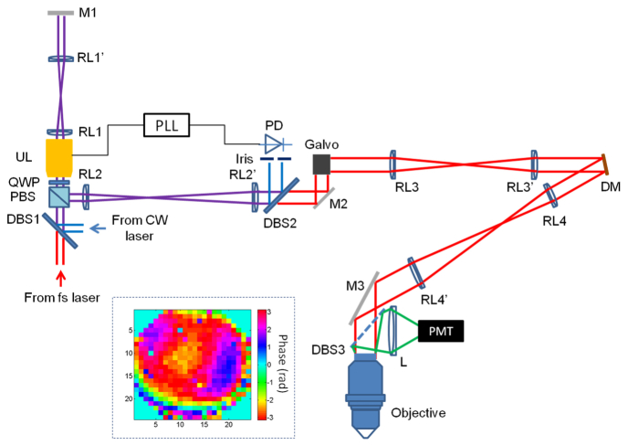
Diagram of the experimental setup. DBS: dichroic beam splitter, PBS: polarization beam splitter, QWP: quarter wave plate, UL: ultrasound lens, RL: relay lens, M: mirror, PLL: phase-lock loop, PD: photodetector, DM: deformable mirror, L: lens, PMT: photomultiplier tube. The inset: phase pattern for correcting the system aberration.
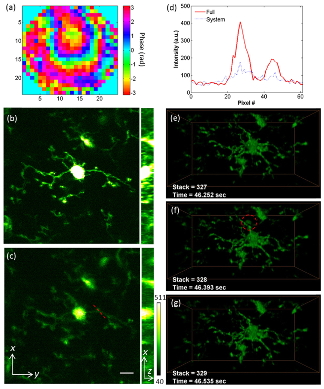
Volumetric imaging of transient morphology of microglia in mouse brain cortex. (a) The phase pattern for full correction of both system and tissue induced wavefront distortions. (b, c) The maximum intensity projections of the microglia at depth 405-413 µm, with full correction and system correction respectively (Visualization 1 ). Scale bar: 10 µm. Power: 36 mW at 935 nm. (d) The signal intensity along the dashed line labeled in (c). (e-g) The transient morphologies of the microglia (Visualization 2 ) at depth 380-420 µm under the dura. Volume size: 98 × 49 × 40 µm3. The dashed circle in (f) labels a GFP-expressing cell patrolling around the brain cortex through a blood vessel. Laser power: 108 mW at 935 nm.
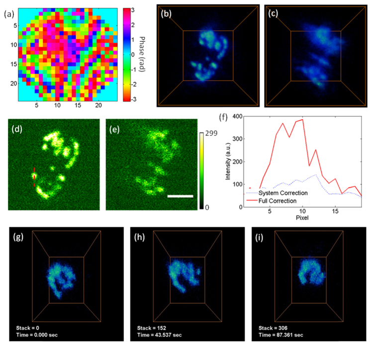
Volumetric imaging of the dynamics of mitochondrial network in lymphocytes of mouse lymph node. (a) The phase pattern for full correction of both system and tissue induced wavefront distortions. (b, c) The volume views of mitochondria in lymphocytes, with full correction and system correction respectively. The volume is 17.6 × 17.6 × 18 µm3, at the depth of 340-364 µm under the surface. Laser power: 55 mW at 935 nm. (d, e) The images acquired at 348 µm depth, with full correction and system correction respectively. Scale bar: 5 µm. (f) The signal intensity along the dashed line labeled in (e). (g-i) The transient morphologies of the mitochondria in lymphocytes (Visualization 3 ). Volume size: 12 × 9 × 18 µm3. Laser power: 90 mW at 935 nm.
Similar articles
-
In vivo fluorescence microscopy via iterative multi-photon adaptive compensation technique.Opt Express. 2014 Oct 6;22(20):23786-94. doi: 10.1364/OE.22.023786. Opt Express. 2014. PMID: 25321957
-
High-resolution in vivo imaging of mouse brain through the intact skull.Proc Natl Acad Sci U S A. 2015 Jul 28;112(30):9236-41. doi: 10.1073/pnas.1505939112. Epub 2015 Jul 13. Proc Natl Acad Sci U S A. 2015. PMID: 26170286 Free PMC article.
-
Superpenetration optical microscopy by iterative multiphoton adaptive compensation technique.Proc Natl Acad Sci U S A. 2012 May 29;109(22):8434-9. doi: 10.1073/pnas.1119590109. Epub 2012 May 14. Proc Natl Acad Sci U S A. 2012. PMID: 22586078 Free PMC article.
-
Advances in adaptive optics-based two-photon fluorescence microscopy for brain imaging.Lasers Med Sci. 2020 Mar;35(2):317-328. doi: 10.1007/s10103-019-02908-z. Epub 2019 Nov 15. Lasers Med Sci. 2020. PMID: 31729608 Review.
-
Biophotonics techniques for structural and functional imaging, in vivo.Anal Cell Pathol (Amst). 2012;35(5-6):317-37. doi: 10.3233/ACP-2012-0058. Anal Cell Pathol (Amst). 2012. PMID: 22433452 Free PMC article. Review.
Cited by
-
Wavefront Shaping Concepts for Application in Optical Coherence Tomography-A Review.Sensors (Basel). 2020 Dec 9;20(24):7044. doi: 10.3390/s20247044. Sensors (Basel). 2020. PMID: 33316998 Free PMC article. Review.
-
The State of the NIH BRAIN Initiative.J Neurosci. 2018 Jul 18;38(29):6427-6438. doi: 10.1523/JNEUROSCI.3174-17.2018. Epub 2018 Jun 19. J Neurosci. 2018. PMID: 29921715 Free PMC article.
-
Mechanical plasticity during oligodendrocyte differentiation and myelination.Glia. 2018 Jan;66(1):5-14. doi: 10.1002/glia.23206. Epub 2017 Sep 21. Glia. 2018. PMID: 28940651 Free PMC article. Review.
-
Multicolor multiphoton in vivo imaging flow cytometry.Opt Express. 2016 Mar 21;24(6):6126-35. doi: 10.1364/OE.24.006126. Opt Express. 2016. PMID: 27136806 Free PMC article.
-
Enhance the delivery of light energy ultra-deep into turbid medium by controlling multiple scattering photons to travel in open channels.Light Sci Appl. 2022 Apr 24;11(1):108. doi: 10.1038/s41377-022-00795-8. Light Sci Appl. 2022. PMID: 35462570 Free PMC article.
References
Publication types
MeSH terms
Grants and funding
LinkOut - more resources
Full Text Sources
Other Literature Sources
Miscellaneous
