

Immunoglobulin G Expression in Human Sperm and Possible Functional Significance
- PMID: 26833114
- PMCID: PMC4735602
- DOI: 10.1038/srep20166
Immunoglobulin G Expression in Human Sperm and Possible Functional Significance
Abstract
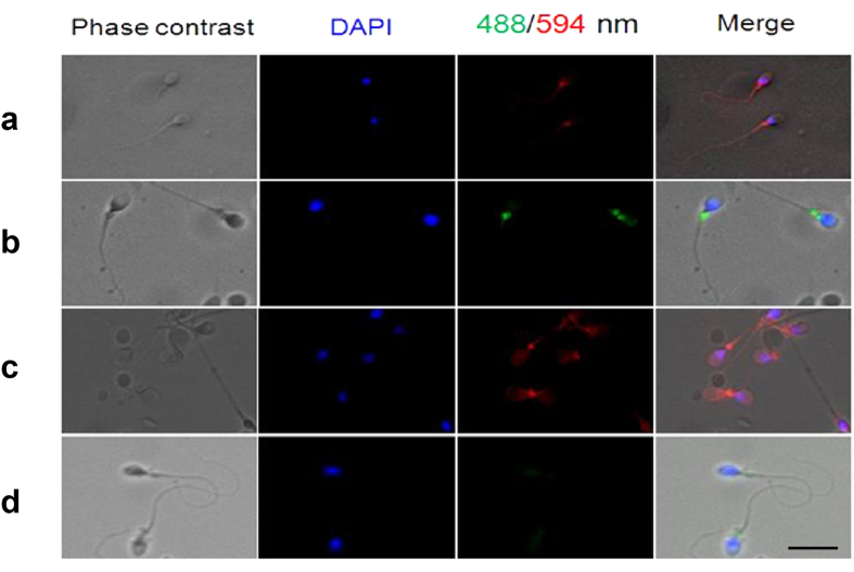
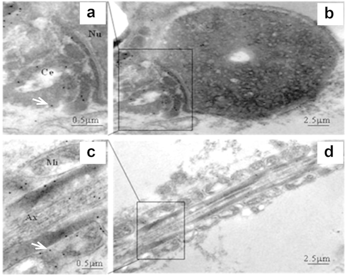
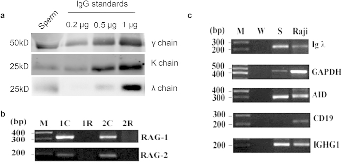
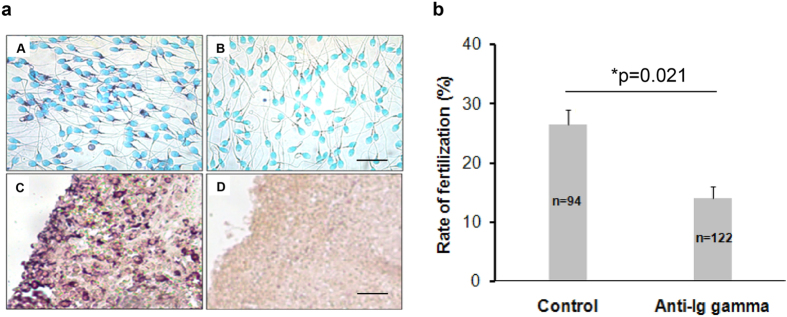
Immunoglobulin G (IgG), the major molecule of the immune system, which was traditionally thought to be produced by differentiated B-lymphocytes, had recently been found in non-immune cells including spermatozoa of rabbit testis. To study if human sperms could produce IgG that might play a role in fertilization, we employed immunofluorescent staining, Western blot, in situ hybridization, RT-PCR (reverse transcription polymerase chain reaction) and immunoelectron microscope and found that human sperms were capable of synthesizing IgG. IgG protein and mRNA were detected in the cytoplasm, mainly the neck region of the sperm and IgG immunoreactivity was found to cover the entire sperm cell. The essential enzymes necessary for IgG synthesis and class switching, RAG1 (recombination activating gene 1), RAG2 (recombination activating gene 2) and AID (activation-induced cytidine deaminase), were also detected in the sperm cells. Furthermore, we found that anti-IgG antibody could inhibit sperm from penetrating Zona-free hamster egg with statistical significance. These discoveries suggested that immunoglobulin G could be produced by human sperms and it might play a role during fertilization.
Figures
Similar articles
-
Immunoglobulin G expression in carcinomas and cancer cell lines.FASEB J. 2007 Sep;21(11):2931-8. doi: 10.1096/fj.07-8073com. Epub 2007 May 2. FASEB J. 2007. PMID: 17475920
-
Investigation of sperm receptors in the hamster zona pellucida by using univalent (Fab) antibodies to hamster ovary.J Reprod Fertil. 1981 Mar;61(2):257-64. doi: 10.1530/jrf.0.0610257. J Reprod Fertil. 1981. PMID: 7009852
-
Overexpression of immunoglobulin G prompts cell proliferation and inhibits cell apoptosis in human urothelial carcinoma.Tumour Biol. 2013 Jun;34(3):1783-91. doi: 10.1007/s13277-013-0717-z. Epub 2013 Mar 13. Tumour Biol. 2013. PMID: 23483488 Free PMC article.
-
Immunoglobulin g (IgG) expression in human umbilical cord endothelial cells.J Histochem Cytochem. 2011 May;59(5):474-88. doi: 10.1369/0022155411400871. Epub 2011 Mar 23. J Histochem Cytochem. 2011. PMID: 21430258 Free PMC article.
-
Immunological aspects of sperm receptors on the zona pellucida of mammalian eggs.Immunol Commun. 1976;5(5):375-85. doi: 10.3109/08820137609033855. Immunol Commun. 1976. PMID: 61169 Review.
Cited by
-
Sialylated IgG expressed in triple-negative breast cancer cells promotes cancer progression by promoting glycolysis.Breast Cancer Res. 2025 Jun 2;27(1):96. doi: 10.1186/s13058-025-02052-3. Breast Cancer Res. 2025. PMID: 40457370 Free PMC article.
-
Relative Abundance of Spermadhesin-1 in the Seminal Plasma of Young Nellore Bulls Is in Agreement with Reproductive Parameters.Vet Sci. 2023 Oct 7;10(10):610. doi: 10.3390/vetsci10100610. Vet Sci. 2023. PMID: 37888562 Free PMC article.
-
Immunization Elicits Antigen-Specific Antibody Sequestration in Dorsal Root Ganglia Sensory Neurons.Front Immunol. 2018 Apr 16;9:638. doi: 10.3389/fimmu.2018.00638. eCollection 2018. Front Immunol. 2018. PMID: 29755449 Free PMC article.
-
The Expression of Non B Cell-Derived Immunoglobulins.Adv Exp Med Biol. 2024;1445:11-36. doi: 10.1007/978-981-97-0511-5_2. Adv Exp Med Biol. 2024. PMID: 38967747 Review.
-
Hepatocyte-derived Igκ promotes HCC progression by stabilizing electron transfer flavoprotein subunit α to facilitate fatty acid β-oxidation.J Exp Clin Cancer Res. 2024 Oct 9;43(1):280. doi: 10.1186/s13046-024-03203-8. J Exp Clin Cancer Res. 2024. PMID: 39380077 Free PMC article.
References
Publication types
MeSH terms
Substances
LinkOut - more resources
Full Text Sources
Other Literature Sources
Molecular Biology Databases
