. 2016 Mar 1;76(5):999-1008.
doi: 10.1158/0008-5472.CAN-15-1439. Epub 2016 Feb 1.

STK11/LKB1 Deficiency Promotes Neutrophil Recruitment and Proinflammatory Cytokine Production to Suppress T-cell Activity in the Lung Tumor Microenvironment

Affiliations

STK11/LKB1 Deficiency Promotes Neutrophil Recruitment and Proinflammatory Cytokine Production to Suppress T-cell Activity in the Lung Tumor Microenvironment

Shohei Koyama et al. Cancer Res. .

Abstract

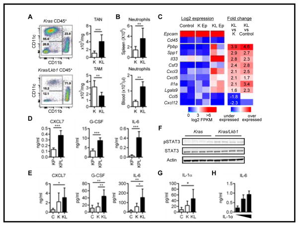
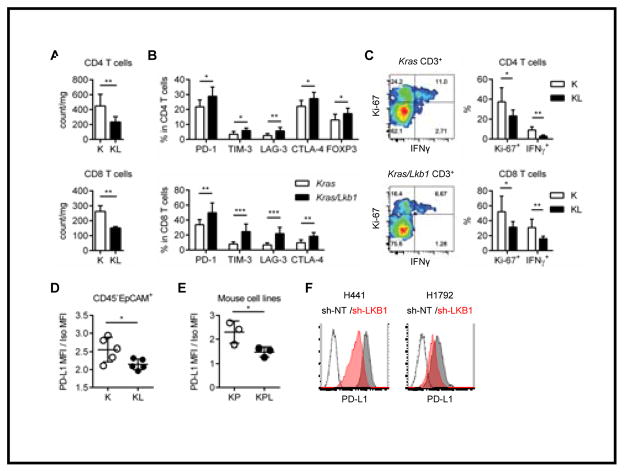
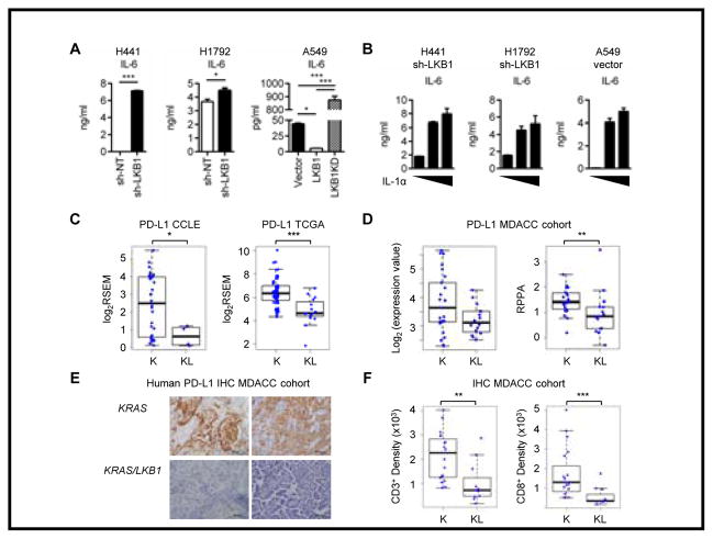
STK11/LKB1 is among the most commonly inactivated tumor suppressors in non-small cell lung cancer (NSCLC), especially in tumors harboring KRAS mutations. Many oncogenes promote immune escape, undermining the effectiveness of immunotherapies, but it is unclear whether the inactivation of tumor suppressor genes, such as STK11/LKB1, exerts similar effects. In this study, we investigated the consequences of STK11/LKB1 loss on the immune microenvironment in a mouse model of KRAS-driven NSCLC. Genetic ablation of STK11/LKB1 resulted in accumulation of neutrophils with T-cell-suppressive effects, along with a corresponding increase in the expression of T-cell exhaustion markers and tumor-promoting cytokines. The number of tumor-infiltrating lymphocytes was also reduced in LKB1-deficient mouse and human tumors. Furthermore, STK11/LKB1-inactivating mutations were associated with reduced expression of PD-1 ligand PD-L1 in mouse and patient tumors as well as in tumor-derived cell lines. Consistent with these results, PD-1-targeting antibodies were ineffective against Lkb1-deficient tumors. In contrast, treating Lkb1-deficient mice with an IL6-neutralizing antibody or a neutrophil-depleting antibody yielded therapeutic benefits associated with reduced neutrophil accumulation and proinflammatory cytokine expression. Our findings illustrate how tumor suppressor mutations can modulate the immune milieu of the tumor microenvironment, and they offer specific implications for addressing STK11/LKB1-mutated tumors with PD-1-targeting antibody therapies.


Conflict of interest statement

Disclosure of Potential Conflicts of Interest:

G.D. received sponsored research support from Bristol-Myers Squibb and Novartis, and is currently an employee of Novartis. He is currently an employee of Novartis. G.J.F. receives patent royalties on the PD-1 pathway from Bristol-Myers-Squibb, Roche, Merck, EMD-Serrono, Boehringer-Ingelheim, Amplimmune/AstraZeneca, and Novartis. F.S.H. is a Bristol-Myers Squibb nonpaid consultant, Novartis, Merck and Genentech consultant and receives clinical trial support to the institution from these companies.

Figures

Figure 1
Figure 1. Tumor-suppressor Lkb1 inactivation promotes neutrophil accumulation via proinflammatory cytokines and chemokines
A. Immune cell populations in the lung tumors from Kras (K) and Kras/Lkb1 (KL) mouse models. Representative flow cytometry data (live/single/total CD45+ cells) from each mouse model (left). Total counts of tumor associated neutrophils (TAN): CD11b+Ly-6G+ cells and tumor associated macrophages (TAM): CD11c+CD11bCD103 from K (n=8) and KL (n=8) mice. **p<0.01, ***p<0.001. B. Neutrophil counts in the spleen and peripheral blood from K (n=8) or KL (n=8) mice (right). **p<0.01. C. Expression of immune modulating factors from RNA sequencing of the sorted tumor cells (CD45EpCAM+) in Kras (K Ep) or Kras/Lkb1 (KL Ep) mice and uninduced normal lung CD45EpCAM+ cells (Control). Each column consists of a combination of samples derived from 3–4 mice. Log-transformed FPKM values are shown, colored blue/red for low/high expression, respectively. Epcam and Cd45 expression are shown as positive and negative controls. Differential expression is shown as fold-change values, colored blue/red for under/over-expression compared to controls. D, E. Chemokine and cytokine levels in the culture supernatants after 48hr incubation from Kras/p53 (KP) (n=3) versus Kras/p53/Lkb1 (KPL) (n=3) cell lines generated from mouse lung tumors, ***p<0.001. Data indicate three replicate wells and are representative of three independent experiments (D) and bronchoalveolar lavage fluid (BALF)s from littermate controls (n=5), K mice (n=8) or KL mice (n=8). *p<0.05, **p<0.01 (E). F. Western blot analysis for pSTAT3, STAT3 levels in K versus KL tumors. Each column represents tumor from a different mouse and actin represents loading control. G. IL-1α level in the BALF from C (n=5), K (n=8) or KL (n=8) mice. *p<0.05. H. IL-6 levels in culture supernatants measured 24hr after IL-1α stimulation (0, 5 and 20ng/ml) of KP (n=3) versus KPL (n=3) cell lines. Data indicate three replicate wells and are representative of three independent experiments.
Figure 2
Figure 2. Lkb1 inactivation leads to a T cell suppressive tumor microenvironment with low PD-L1 expression in tumor cells
A. Total counts of CD4 T cells (top) and CD8 T cells (bottom). **p<0.01. B. Expression of checkpoint receptors in CD4 T cells (top) and CD8 T cells (bottom) in K or KL tumors. *p<0.05, **p<0.01, ***p<0.001. C. IFNγ expression and proliferation marker (Ki-67) positivity for CD4 or CD8 T cells in K or KL tumors. Representative flow cytometry data (total CD3+ T cells) from each mouse model (left). Percentage of Ki-67+ and IFNγ+ in CD4 or CD8 T cells from K (n=6) or KL (n=6) mice. *p<0.05, **p<0.01. D. PD-L1 expression in gated CD45EpCAM+ cells in K (n=5) or KL (n=5) tumors evaluated by flow cytometry. *p=0.0384. E. PD-L1 expression in KP (n=3) versus KPL (n=3) cell lines evaluated by flow cytometry. *p=0.0495. Data is representative of three independent experiments. F. PD-L1 expression in H441 or H1792 cells stably transfected with sh-non-target (NT) or sh-LKB1. Data are representative of three independent experiments.
Figure 3
Figure 3. LKB1 inactivation in human KRAS mutated cell lines showed similar phenotype with mouse Kras/Lkb1 tumor
A. Analysis of IL-6 in the culture supernatants after 48hr incubation of KRAS mutated LKB1 wild type H441 or H1792 cells stably transfected with sh-NT or sh-LKB1 and KRAS, LKB1 mutant A549 cells reconstituted with empty vector (Vector), wild type LKB1 or Kinase dead LKB1 (LKB1KD). *p<0.05, ***p<0.001. Data indicate three replicate wells and are representative of three independent experiments. B. IL-6 levels in culture supernatants measured 24hr after IL-1α stimulation (0, 5 and 20ng/ml) of three LKB1-deficient cell lines. Data indicate three replicate wells and are representative of three independent experiments. C. PDL1 expression in KRAS (K) or KRAS and LKB1 mutated (KL) cell lines from CCLE database (*p=0.04) and PDL1 expression in K or KL lung adenocarcinoma samples from TCGA database (***p=0.00004). D. PD-L1 mRNA levels determined by microarray (p=0.1) and protein levels (**p=0.009) determined by RPPA from the MDACC dataset. E. Representative immunohistochemistry for PD-L1 on the KRAS mutated LKB1 wt or mutant patient tumors from the MDACC cohort. F. CD3 (**p=0.002) and CD8 (***p=0.0003) positive cell densities by immunohistochemistry on the MDACC patient cohorts.
Figure 4
Figure 4. IL-6 neutralizing treatment showed clinical efficacy in Kras/Lkb1 mouse model
A. Representative images of Magnetic resonance imaging (MRI) and quantification of MRI from KL mice treated with PD-1 or IL-6 blocking antibodies or controls. B. Survival of untreated mice vs mice treated with IL-6 blocking antibody (***p=0.0002, n=6 vs 12 respectively). C, D. IL-6 and G-CSF levels in BALFs (C) and TAN counts (D) for untreated KL mice (n=7) or KL mice treated with IL-6 neutralizing antibody (n=8) with comparable tumor burden. *p<0.05, **p<0.01. E. Ki-67 and IFNγ positive CD8 T cell counts in untreated KL mice (n=7) or KL mice treated with IL-6 neutralizing antibody (n=8) with comparable tumor burden. F. Representative Ki-67 and TUNEL immunohistochemistry and quantification per the microscopic field on the KL mice untreated or treated with IL-6 neutralizing antibody. Each data point represents a different microscopic field. For Ki-67 n=9 and 5 and for TUNEL n=8 and 5 for untreated and IL-6 ab treated mice respectively. **p=0.0049 for Ki-67 and **p=0.0024 for TUNEL.

References

    1. Cardarella S, Johnson BE. The impact of genomic changes on treatment of lung cancer. American journal of respiratory and critical care medicine. 2013;188(7):770–5. - PMC - PubMed
    1. Comprehensive molecular profiling of lung adenocarcinoma. Nature. 2014;511(7511):543–50. - PMC - PubMed
    1. Ji H, Ramsey MR, Hayes DN, Fan C, McNamara K, Kozlowski P, et al. LKB1 modulates lung cancer differentiation and metastasis. Nature. 2007;448(7155):807–10. - PubMed
    1. Chen Z, Cheng K, Walton Z, Wang Y, Ebi H, Shimamura T, et al. A murine lung cancer co-clinical trial identifies genetic modifiers of therapeutic response. Nature. 2012;483(7391):613–7. - PMC - PubMed
    1. Zhao N, Wilkerson MD, Shah U, Yin X, Wang A, Hayward MC, et al. Alterations of LKB1 and KRAS and risk of brain metastasis: Comprehensive characterization by mutation analysis, copy number, and gene expression in non-small-cell lung carcinoma. Lung cancer (Amsterdam, Netherlands) 2014 - PMC - PubMed

Publication types

MeSH terms