. 2016 Feb 1;198(7):1171-81.
doi: 10.1128/JB.00961-15.

L-Hydroxyproline and d-Proline Catabolism in Sinorhizobium meliloti

Affiliations

L-Hydroxyproline and d-Proline Catabolism in Sinorhizobium meliloti

Siyun Chen et al. J Bacteriol. .

Abstract

Sinorhizobium meliloti forms N2-fixing root nodules on alfalfa, and as a free-living bacterium, it can grow on a very broad range of substrates, including l-proline and several related compounds, such as proline betaine, trans-4-hydroxy-l-proline (trans-4-l-Hyp), and cis-4-hydroxy-d-proline (cis-4-d-Hyp). Fourteen hyp genes are induced upon growth of S. meliloti on trans-4-l-Hyp, and of those, hypMNPQ encodes an ABC-type trans-4-l-Hyp transporter and hypRE encodes an epimerase that converts trans-4-l-Hyp to cis-4-d-Hyp in the bacterial cytoplasm. Here, we present evidence that the HypO, HypD, and HypH proteins catalyze the remaining steps in which cis-4-d-Hyp is converted to α-ketoglutarate. The HypO protein functions as a d-amino acid dehydrogenase, converting cis-4-d-Hyp to Δ(1)-pyrroline-4-hydroxy-2-carboxylate, which is deaminated by HypD to α-ketoglutarate semialdehyde and then converted to α-ketoglutarate by HypH. The crystal structure of HypD revealed it to be a member of the N-acetylneuraminate lyase subfamily of the (α/β)8 protein family and is consistent with the known enzymatic mechanism for other members of the group. It was also shown that S. meliloti can catabolize d-proline as both a carbon and a nitrogen source, that d-proline can complement l-proline auxotrophy, and that the catabolism of d-proline is dependent on the hyp cluster. Transport of d-proline involves the HypMNPQ transporter, following which d-proline is converted to Δ(1)-pyrroline-2-carboxylate (P2C) largely via HypO. The P2C is converted to l-proline through the NADPH-dependent reduction of P2C by the previously uncharacterized HypS protein. Thus, overall, we have now completed detailed genetic and/or biochemical characterization of 9 of the 14 hyp genes.

Importance: Hydroxyproline is abundant in proteins in animal and plant tissues and serves as a carbon and a nitrogen source for bacteria in diverse environments, including the rhizosphere, compost, and the mammalian gut. While the main biochemical features of bacterial hydroxyproline catabolism were elucidated in the 1960s, the genetic and molecular details have only recently been determined. Elucidating the genetics of hydroxyproline catabolism will aid in the annotation of these genes in other genomes and metagenomic libraries. This will facilitate an improved understanding of the importance of this pathway and may assist in determining the prevalence of hydroxyproline in a particular environment.


Figures

FIG 1
FIG 1
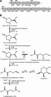
Genetics and biochemistry of hydroxyproline metabolism in S. meliloti. (A) Diagram of the hydroxyproline transport and catabolic locus; (B) schematic diagram of the hydroxyproline catabolic pathway, as described by Adams and Frank (11). (A and B) Gene annotations, promoters, and enzymatic functions as deduced through this study and previous work (13, 14, 18). HypR, negative regulator; HypD, Δ1-pyrroline-4-hydroxy-2-carboxylate deaminase; HypT, unknown; HypS, Δ1-pyrroline-2-carboxylate reductase; HypH, α-ketoglutarate semialdehyde dehydrogenase; HypMNPQ, l-hydroxyproline ABC-type transport system; HypO, cis-4-hydroxy-d-proline dehydrogenase; HypRE, hydroxyproline 2-epimerase; HypX, unknown; HypY, unknown, a possible proline racemase pseudogene; HypZ, unknown.
FIG 2
FIG 2
Schematic representation of the HypD crystal structure. (A) View from the “side” of the (α/β)8 barrel. The barrel motif is to the right of the image, and alpha helices 9, 10, 13, and 14 of the C-terminal extension are labeled (α9, α10, α13, and α14) for reference. The N terminus, starting at A3 in the refined structure, is labeled with an “N.” (B) View “down” the barrel from the C-terminal face into the active site. The conserved active-site residue K184 is shown. All alpha helices are labeled (α1 to α14), as are the beta sheets (β1 to β8) of the (α/β)8 barrel motif. (A and B) Structures are colored from the N terminus (blue) to the C terminus (red).
FIG 3
FIG 3
Growth profiles of S. meliloti hypS and related mutants. Shown are growth profiles of several S. meliloti strains in minimal medium containing as a sole source of carbon l-proline (A), d-proline (B), or trans-4-hydroxy-l-proline (C). Data points are the means from triplicate samples, and the error bars indicate the standard deviations. Strains shown are wild-type RmP110, RmP2514 (ΔhypS), RmP3155 (proC::Tn5-B20 ΔB161), RmP3153 (proC::Tn5-B20 ΔB161 ΔhypS), and RmP3272 [ΔhypS pTH2685(hypS+)].
FIG 4
FIG 4
Induction profiles of the hypM, hypS, and hypR genes. The expression levels of the three hyp genes were measured following 40 h of growth in four different carbon sources using a gusA transcriptional fusion. β-Glucuronidase activities were derived from triplicate assays (±the standard errors of the mean), and similar results were obtained in two independent experiments. Strains shown are wild-type RmP110, RmP1886 (hypM::gusA), RmP239 (hypS::gusA), and RmFL2315 (hypR::gusA).
FIG 5
FIG 5
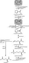
Schematic illustrations of l- and d-proline metabolism in Sinorhizobium meliloti. The biosynthetic and catabolic pathways for l-proline biosynthesis from l-glutamate are shown, as is the proposed pathway for d-proline catabolism. l-Proline and d-proline are highlighted by gray shading. The association of proteins to each biochemical reaction is based on the work reported here and elsewhere (20, 49, 64, 65, 67). Abbreviations: Pyr5C, Δ1-pyrroline-5-carboxylate; Pyr2C, Δ1-pyrroline-2-carboxylate; γ-glutamyl-5-P, γ-glutamyl-5-phosphate; Pi, phosphate.
FIG 6
FIG 6
Effect of a putA mutation on growth with proline compounds. The growth profiles of wild-type S. meliloti RmP110 and S. meliloti RmFL5502 (putA::pTH1522) are shown in minimal medium containing as a sole source of carbon l-proline (A), d-proline (B), or trans-4-hydroxy-l-proline (C). Data points are the means from triplicate samples, and the error bars indicate the standard deviations.

References

    1. Lawrence CC, Sobey WJ, Field RA, Baldwin JE, Schofield CJ. 1996. Purification and initial characterization of proline 4-hydroxylase from Streptomyces griseoviridis P8648: a 2-oxoacid, ferrous-dependent dioxygenase involved in etamycin biosynthesis. Biochem J 313(Part 1):185–191. doi:10.1042/bj3130185. - DOI - PMC - PubMed
    1. Shibasaki T, Mori H, Chiba S, Ozaki A. 1999. Microbial proline 4-hydroxylase screening and gene cloning. Appl Environ Microbiol 65:4028–4031. - PMC - PubMed
    1. Gorres KL, Raines RT. 2010. Prolyl 4-hydroxylase. Crit Rev Biochem Mol Biol 45:106–124. doi:10.3109/10409231003627991. - DOI - PMC - PubMed
    1. Lamport DT, Miller DH. 1971. Hydroxyproline arabinosides in the plant kingdom. Plant Physiol 48:454–456. doi:10.1104/pp.48.4.454. - DOI - PMC - PubMed
    1. Kieliszewski MJ, O'Neill M, Leykam J, Orlando R. 1995. Tandem mass spectrometry and structural elucidation of glycopeptides from a hydroxyproline-rich plant cell wall glycoprotein indicate that contiguous hydroxyproline residues are the major sites of hydroxyproline O-arabinosylation. J Biol Chem 270:2541–2549. doi:10.1074/jbc.270.6.2541. - DOI - PubMed

Publication types

MeSH terms

LinkOut - more resources