Review
. 2016 Mar 31;198(8):1186-95.
doi: 10.1128/JB.00901-15. Print 2016 Apr.

Minicells, Back in Fashion

Affiliations
Review

Minicells, Back in Fashion

Madeline M Farley et al. J Bacteriol. .

Abstract

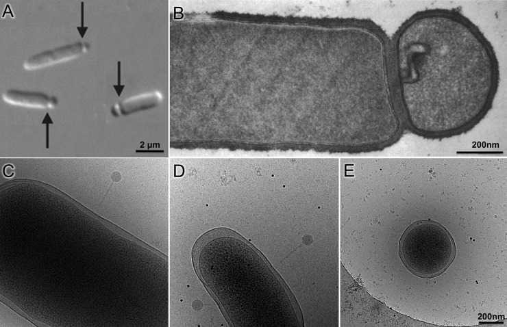
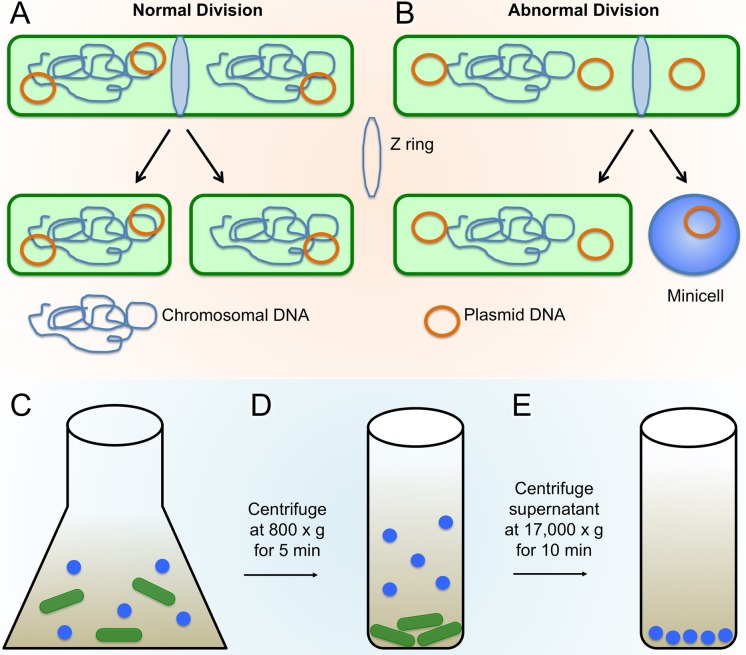
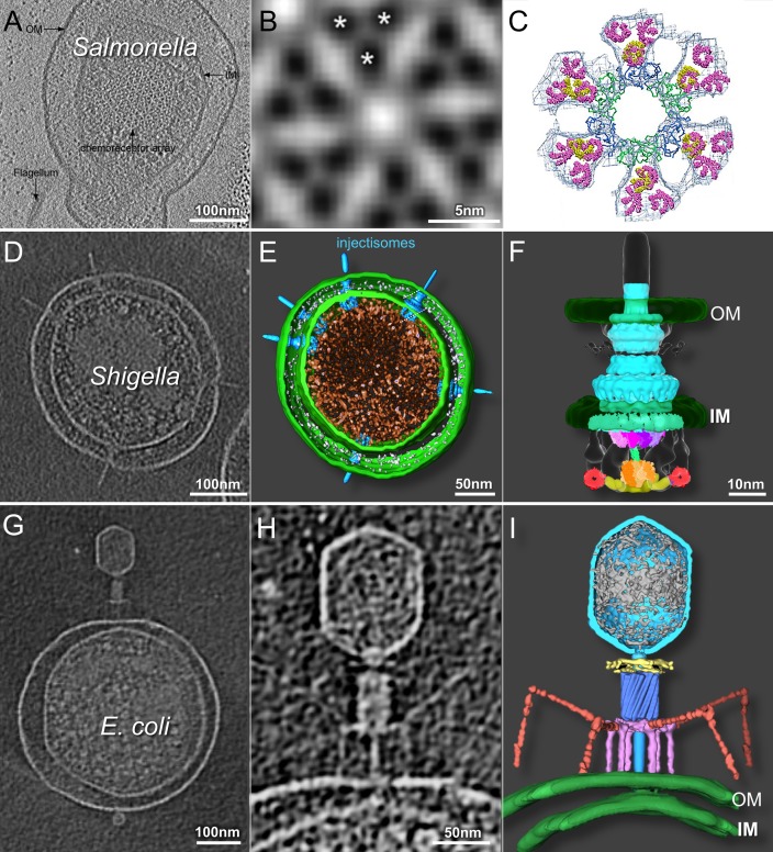
Cryo-electron tomography (cryo-ET) has emerged as a leading technique for three-dimensional visualization of large macromolecular complexes and their conformational changes in their native cellular environment. However, the resolution and potential applications of cryo-ET are fundamentally limited by specimen thickness, preventing high-resolution in situ visualization of macromolecular structures in many bacteria (such as Escherichia coli and Salmonella enterica). Minicells, which were discovered nearly 50 years ago, have recently been exploited as model systems to visualize molecular machines in situ, due to their smaller size and other unique properties. In this review, we discuss strategies for producing minicells and highlight their use in the study of chemotactic signaling, protein secretion, and DNA translocation. In combination with powerful genetic tools and advanced imaging techniques, minicells provide a springboard for in-depth structural studies of bacterial macromolecular complexes in situ and therefore offer a unique approach for gaining novel structural insights into many important processes in microbiology.


Figures

FIG 1
FIG 1
Bacterial minicell production and comparison of minicells to parent cells. (A) Composite differential interference contrast image of minicell formation through polar division in E. coli. (B) Thin-sectioned negative-stain micrograph of B. subtilis minicell (adapted from reference with permission of the publisher). (C to E) Micrographs of cryopreserved wild-type E. coli (C), skinny E. coli (D), and a minicell (E) generated from the skinny E. coli (republished from reference with permission of the publisher). Phage P1 is visible attached to cells in panels C and D. The scale of panels C and D is the same as that in panel E.
FIG 2
FIG 2
Minicell production and enrichment. (A and B) Normal bacterial cell division (A) compared with abnormal division (B), resulting in minicell generation due to polar Z-ring placement (adapted from reference with permission of the publisher). (C to E) Minicells are separated from typical rod-shaped cells through centrifugation; first, the larger rod-shaped cells are pelleted through a short low-speed spin, and then minicells are pelleted from the resulting supernatant fraction through a longer and higher-speed centrifugation.
FIG 3
FIG 3
Cryo-ET of minicells provides 3D structures of chemoreceptor arrays, injectisomes, and phage-host interactions. (A) Slice-through tomographic reconstruction of an S. enterica minicell showcasing the top-down view of the native chemoreceptor array. (B) Subtomographic-average structure of the receptor array resolving the organization of the trimer of dimers, as indicated by asterisks. (C) EM density map fit with the crystal structures of the individual components (republished from reference with permission of the publisher). (D to F) Central tomographic slice of a Shigella minicell with multiple injectisomes (D), corresponding 3D surface rendering (E), and surface rendering of the injectisome basal body (republished from reference with permission of the publisher) fit with structures of the isolated components (F) (republished from reference with permission of the publisher). (G to I) Tomographic slice through the center of an E. coli minicell infected with phage T4 (G), magnified view of the same adsorbed phage particle penetrating and transferring DNA into the cell (H), and the corresponding 3D rendering (republished from reference with permission of the publisher) (I). OM, outer membrane; IM, inner membrane.

References

    1. Alberts B. 1998. The cell as a collection of protein machines: preparing the next generation of molecular biologists. Cell 92:291–294. doi:10.1016/S0092-8674(00)80922-8. - DOI - PubMed
    1. Lučič V, Rigort A, Baumeister W. 2013. Cryo-electron tomography: the challenge of doing structural biology in situ. J Cell Biol 202:407–419. doi:10.1083/jcb.201304193. - DOI - PMC - PubMed
    1. Dubochet J, Adrian M, Chang JJ, Homo JC, Lepault J, McDowall AW, Schultz P. 1988. Cryo-electron microscopy of vitrified specimens. Q Rev Biophys 21:129–228. doi:10.1017/S0033583500004297. - DOI - PubMed
    1. Cheng Y. 2015. Single-particle cryo-EM at crystallographic resolution. Cell 161:450–457. doi:10.1016/j.cell.2015.03.049. - DOI - PMC - PubMed
    1. Hoenger A. 2014. High-resolution cryo-electron microscopy on macromolecular complexes and cell organelles. Protoplasma 251:417–427. doi:10.1007/s00709-013-0600-1. - DOI - PMC - PubMed

Publication types

MeSH terms

Substances