. 2016 Feb 1;8(2):458-66.
doi: 10.1093/gbe/evw011.

Group II Intron-Mediated Trans-Splicing in the Gene-Rich Mitochondrial Genome of an Enigmatic Eukaryote, Diphylleia rotans

Affiliations

Group II Intron-Mediated Trans-Splicing in the Gene-Rich Mitochondrial Genome of an Enigmatic Eukaryote, Diphylleia rotans

Ryoma Kamikawa et al. Genome Biol Evol. .

Abstract

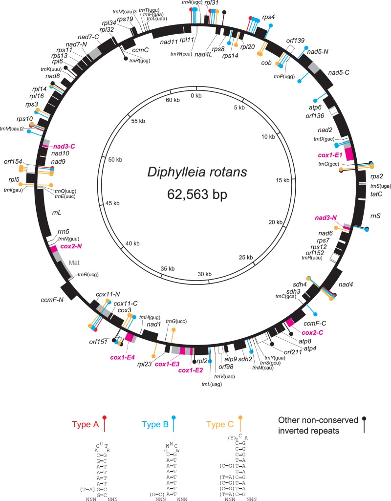
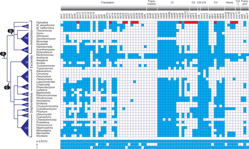
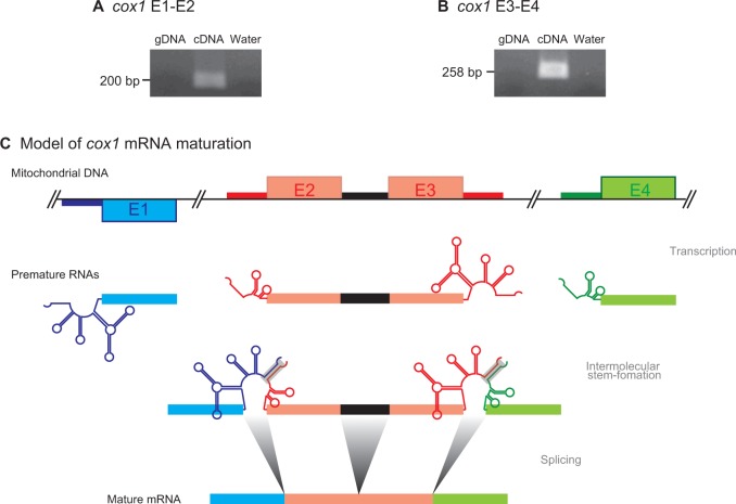
Although mitochondria have evolved from a single endosymbiotic event, present day mitochondria of diverse eukaryotes display a great range of genome structures, content and features. Group I and group II introns are two features that are distributed broadly but patchily in mitochondrial genomes across branches of the tree of eukaryotes. While group I intron-mediated trans-splicing has been reported from some lineages distantly related to each other, findings of group II intron-mediated trans-splicing has been restricted to members of the Chloroplastida. In this study, we found the mitochondrial genome of the unicellular eukaryote Diphylleia rotans possesses currently the second largest gene repertoire. On the basis of a probable phylogenetic position of Diphylleia, which is located within Amorphea, current mosaic gene distribution in Amorphea must invoke parallel gene losses from mitochondrial genomes during evolution. Most notably, although the cytochrome c oxidase subunit (cox) 1 gene was split into four pieces which located at a distance to each other, we confirmed that a single mature mRNA that covered the entire coding region could be generated by group II intron-mediated trans-splicing. This is the first example of group II intron-mediated trans-splicing outside Chloroplastida. Similar trans-splicing mechanisms likely work for bipartitely split cox2 and nad3 genes to generate single mature mRNAs. We finally discuss origin and evolution of this type of trans-splicing in D. rotans as well as in eukaryotes.

Keywords: Diphyllatia; introns; inverted repeats; mitochondria; trans-splicing.


Figures

F<sc>ig</sc>. 1.—
Fig. 1.—
The complete mitochondrial genome of D. rotans. Functionally identifiable protein-coding, SSU rRNA, and LSU rRNA genes are depicted as closed boxes whereas unidentified ORFs are as open boxes. 5S rRNA and tRNA genes are shown as lines. Intron sequences are colored in gray. Split genes are highlighted in magenta. Three types of conserved palindromic sequences and nonconserved palindromic sequences are indicated by different colors: type A in red, type B in blue, type C in yellow, and nonconserved type in black.
F<sc>ig</sc>. 2.—
Fig. 2.—
Distribution of protein coding genes in mitochondrial genomes. Presence and absence of corresponding genes in mitochondrial genomes of various eukaryotes is shown by closed and open boxes, respectively. The gene contents were determined from genome sequences retrieved from the GenBank. Rare genes found in the D. rotans mitochondrial genome are highlighted in red. Phylogenetic relationships of eukaryotes are based on Derelle et al. (2015), Brown et al. (2013), and Eme et al. (2014). The predicted protein gene contents of LECA (a), the last common ancestor of Amorphea (b), and that of Diapholetickes and Excavata (c), are shown. Ma: Malawimonas; Op: Opisthokonta; Am: Amoebozoa; Di: Discoba; Al: Alveolata; St: Stramenopiles; Rh: Rhizaria; Cr: Cryptophyceae; Ha: Haptophyta; Re: Red algae (Rhodophyceae); Gl: Glaucophyta; Ch: Chloroplastida; CI–CV: electron transport chain complex I–V.
F<sc>ig</sc>. 3.—
Fig. 3.—
Trans-splicing in cox1 transcripts. (A) Reverse transcriptase PCR for transcripts of cox1-E1 and cox1-E2. Genomic DNA (left lane), cDNA (middle lane), and distilled water (right lane) were used as templates. (B) Reverse transcriptase PCR for transcripts of cox1-E3 and cox1-E4. Genomic DNA (left lane), cDNA (middle lane), and distilled water (right lane) were used as templates. (C) A model of cox1 mRNA maturation. Mitochondrial DNA, cox1 coding regions, and intron regions are depicted as thin lines, boxes, and thick lines, respectively. Independent gene fragments and their transcripts are distinguished by different colors, whereas the group I intron splitting cox1-E2 and cox1-E3 is colored in black. Four cox1 gene fragments are located on the mitochondrial DNA separately. Cox1-E2 and cox1-E3 are separated by insertion of a cis-spliced group I intron. In this model, premature RNAs from separately distributed gene fragments for cox1 are transcribed with flanking intron regions. Flanking intron regions form intermolecular stem structures that associated to form group II intron secondary structures.

Similar articles

Cited by

References

    1. Adams KL, Palmer JD. 2003. Evolution of mitochondrial gene content: gene loss and transfer to the nucleus. Mol Phylogenet Evol. 29:380–395. - PubMed
    1. Adl SM, et al. 2012. The revised classification of eukaryotes. J Eukaryot Microbiol. 59:429–493. - PMC - PubMed
    1. Altschul SF, Gish W, Miller W, Myers EW, Lipman DJ. 1990. Basic local alignment search tool. J Mol Biol. 215:403–410. - PubMed
    1. Beaudet D, et al. 2013. Mitochondrial genome rearrangements in Glomus species triggered by homologous recombination between distinct mtDNA haplotypes. Genome Biol Evol. 5:1628–1643. - PMC - PubMed
    1. Brown MW, et al. 2013. Phylogenomics demonstrates that breviate flagellates are related to opisthokonts and apusomonads. Proc Biol Sci. 280:20131755. - PMC - PubMed

Publication types

Substances