. 2016 Apr 8;291(15):8295-307.
doi: 10.1074/jbc.M116.714568. Epub 2016 Feb 1.

Multiple Domains of GlcNAc-1-phosphotransferase Mediate Recognition of Lysosomal Enzymes

Affiliations

Multiple Domains of GlcNAc-1-phosphotransferase Mediate Recognition of Lysosomal Enzymes

Eline van Meel et al. J Biol Chem. .

Abstract

The Golgi enzyme UDP-GlcNAc:lysosomal enzymeN-acetylglucosamine-1-phosphotransferase (GlcNAc-1-phosphotransferase), an α2β2γ2hexamer, mediates the initial step in the addition of the mannose 6-phosphate targeting signal on newly synthesized lysosomal enzymes. This tag serves to direct the lysosomal enzymes to lysosomes. A key property of GlcNAc-1-phosphotransferase is its unique ability to distinguish the 60 or so lysosomal enzymes from the numerous non-lysosomal glycoproteins with identical Asn-linked glycans. In this study, we demonstrate that the two Notch repeat modules and the DNA methyltransferase-associated protein interaction domain of the α subunit are key components of this recognition process. Importantly, different combinations of these domains are involved in binding to individual lysosomal enzymes. This study also identifies the γ-binding site on the α subunit and demonstrates that in the majority of instances the mannose 6-phosphate receptor homology domain of the γ subunit is required for optimal phosphorylation. These findings serve to explain how GlcNAc-1-phosphotransferase recognizes a large number of proteins that lack a common structural motif.

Keywords: lysosomal glycoprotein; lysosomal storage disease; lysosome; mannose 6-phosphate (Man-6-P); phosphorylation.


Figures

FIGURE 1.
FIGURE 1.
Generation of GNPTAB−/− and GNPTG−/− HeLa cell lines using CRISPR/Cas9 genome editing. A and B, schematic of the base pairing between a guide RNA (sgRNA_g11 for GNPTAB and sgRNA_g8 for GNPTG) and the targeting loci of the two genes. The sequences of the mutated alleles are shown. All the insertions and deletions cause frameshift mutations and early termination of translation.
FIGURE 2.
FIGURE 2.
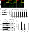
GNPTAB−/− and GNPTG−/− HeLa cells have enlarged lysosomes and are deficient in phosphorylated lysosomal proteins. A, confocal immunofluorescence images of parental, α/β-deficient, and γ-deficient HeLa cells, stained for the late endosomal/lysosomal marker LAMP-1 (green). Scale bar, 10 μm. B, immunoblot of cell lysates (20 μg per lane) from parental, α/β-deficient, and γ-deficient HeLa cells probed for LAMP-1. C, lysates of the three cell lines were incubated with CI-MPR affinity beads, and the binding of the various lysosomal proteins was determined by enzyme assays. Mean values obtained with the parental HeLa cells are set to 100% ± S.D. (n = 3–5). D, lysates of the three cell lines were incubated with CI-MPR affinity beads, and the binding of the three lysosomal proteins was determined by Western blots. E, WT and mutant cells transfected with plasmids encoding the four lysosomal proteins were labeled with [2-3H]mannose, followed by immunoprecipitation of the proteins secreted into the media and determination of the percent N-glycans containing Man-6-P. The percentage oligosaccharide phosphorylation ± S.D. for each enzyme in the three different cell lines is shown (n = 2–5).
FIGURE 3.
FIGURE 3.
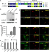
The ΔN1-DMAP mutant has catalytic activity but is unable to phosphorylate lysosomal enzymes. A, schematic of GlcNAc-1-phosphotransferase α/β subunit modular arrangement and deletion of the Notch 1 through DMAP interaction domain (residues 438–819). B, immunoblot analysis of WT α/β versus the ΔN1-DMAP deletion mutant expressed in GNPTAB−/− HeLa cells probed with anti-V5 antibody. C, confocal immunofluorescence images of parental HeLa cells transfected with WT α/β or the ΔN1-DMAP mutant and co-localized with the Golgi markers GM130 or GOLPH4, respectively (see “Experimental Procedures”). D, phosphotransferase activity toward the simple sugar αMM, using extracts of GNPTAB−/− cells transfected with vector, WT α/β, or ΔN1-DMAP mutant plasmids. E, transfection of GNPTAB−/− HeLa cells with WT α/β but not the ΔN1-DMAP mutant restores lysosomal enzyme phosphorylation as determined by binding to CI-MPR affinity beads. Mean values obtained with cells transfected with WT α/β are set to 100% ± S.D. F, confocal immunofluorescence images of GNPTAB−/− cells co-expressing γ with WT α/β (top) or with the ΔN1-DMAP mutant (bottom). Scale bars, 10 μm.
FIGURE 4.
FIGURE 4.
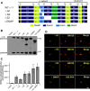
Expression of deletion mutants of α/β. A, schematic of the various α/β deletion constructs expressed in HEK 293 cells. B, Western blot of WT α/β and mutants expressed in GNPTAB−/− HeLa cells. 20 μg of each cell extract was loaded, and the α/β precursor and β subunits were detected with an anti-V5 antibody. C, catalytic activity of WT α/β and the mutants toward αMM using equal amounts of whole cell extracts. The vector-only transfected GNPTAB−/− HeLa cell extract served as a control. D, confocal immunofluorescence microscopy of HeLa cells transfected with plasmids encoding the various α/β deletion mutants. Cells were fixed 24 h post-transfection and probed with anti-α antibody (ΔN1, ΔN2, and ΔS2), or with the anti-V5 antibody (ΔDMAP). The Golgi markers, GM130 and GOLPH4, were detected with anti-GM130 and anti-GOLPH4 antibodies, respectively. Scale bar, 10 μm.
FIGURE 5.
FIGURE 5.
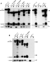
The γ subunit binds to amino acids 535–694 of the α subunit. A and B, lysates of HEK 293 cells expressing WT α/β or the deletion mutants along with WT γ were incubated with Ni-NTA affinity beads to pull down the various forms of the α/β subunits. γ only served as a control. The bound material was assayed for the content of α/β and γ subunits by SDS-PAGE and Western blotting using anti-V5 antibody to detect α/β and anti-HA antibody (A) or anti-FLAG antibody (B) to detect γ. I, 3% of input; P, 25% of pellet fraction. *, line denotes position where the nitrocellulose membrane was cut to allow for probing of the α/β and γ subunits with different antibodies. Even long exposures of the blot failed to show detectable binding of γ to the ΔN1-DMAP, ΔN2-DMAP, and ΔS2 mutants of α/β (data not shown).
FIGURE 6.
FIGURE 6.
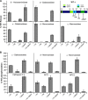
Effect of the α/β domain deletions on lysosomal enzyme phosphorylation. A, lysates of GNPTAB−/− HeLa cells transfected with WT or deletion mutants of α/β were incubated with CI-MPR affinity beads, and the bound material was assayed for the content of the various acid glycosidases. Mean values obtained with cells transfected with the various mutants are compared with WT α/β, which is set to 100% (n = 3–5). B, GNPTAB−/− cells were co-transfected with the various forms of α/β along with the indicated lysosomal protein encoding plasmids. Following [2-3H]mannose labeling, the degree of N-glycan phosphorylation of the lysosomal proteins was determined. Values obtained with the various mutants are compared with WT α/β, which is set to 100% (n = 2–3).
FIGURE 7.
FIGURE 7.
Effect of substituting the Notch 2 repeat module with Notch 1. A, schematic showing α/β construct with two Notch 1 modules. B, Western blot of GNPTAB−/− cells transfected with either WT α/β, the 2Notch1 mutant, or the ΔN2 mutant. 20 μg of each cell lysate was loaded. C, confocal immunofluorescence images of GNPTAB−/− cells expressing the 2Notch1 mutant. Cells were fixed 24 h post-transfection and probed with anti-α antibody (2 Notch 1) and anti-GM130 antibody for the Golgi marker GM130. Scale bar, 10 μm. D, lysates of HEK 293 cells expressing WT α/β or the 2Notch1 mutant along with WT γ were incubated with Ni-NTA affinity beads to pull down the various forms of the α/β subunits. γ only served as a control. The bound material was assayed for the content of α/β and γ subunits by SDS-PAGE and Western blotting using anti-V5 antibody to detect α/β and anti-FLAG antibody to detect γ. I, 3% of input; P, 25% of pellet fraction. E, phosphotransferase activity toward the simple sugar αMM, using extracts of GNPTAB−/− cells transfected with either vector, WT α/β, the 2Notch1 mutant, or the ΔN2 mutant. n = 3. F, GNPTAB−/− HeLa cells transfected with either WT α/β, the 2Notch1 mutant, or the ΔN2 mutant were assayed for activity of the indicated enzymes as determined by binding to CI-MPR affinity beads. Mean values obtained with the two mutants are compared with WT α/β, which is set to 100% ± S.D. (n = 3). *, line denotes position where the nitrocellulose membrane was cut to allow for probing of the α/β and γ subunits with different antibodies.
FIGURE 8.
FIGURE 8.
The γDMAP interaction domain binds lysosomal enzymes. A, human α/β and γDMAP interaction domains are aligned with identical amino acid residues shown in red. B, alignment of the DMAP interaction domains of γ from several species shows a high degree of conservation in the N-terminal half of the domain. Because of the variation of 5 amino acid residues, only bovine γ shows up as a DMAP interaction domain in an NCBI Blast search. C, pulldown assays were performed with GST fusions to α/β and γDMAP interaction domains using the purified lysosomal enzymes indicated, along with tissue factor pathway inhibitor (TFPI), a non-lysosomal secreted glycoprotein. 0.2% of the input and 20% of the pellet fraction was loaded. D, release of bound enzyme activity was measured following wash steps 1 and 2 to determine loss of bound material as a result of the wash steps (n = 2).
FIGURE 9.
FIGURE 9.
Effect of mutating two critical residues in the γMRH domain on lysosomal enzyme phosphorylation. A, schematic showing the domain boundaries of the γ subunit and the position of the MRH mutations. B, confocal immunofluorescence images of GNPTG−/− cells co-transfected with WT α/β and either WT γ or the R134K/E153Q MRH mutant. Scale bar, 10 μm. C, lysates of HEK 293 cells expressing WT α/β along with WT γ or the R134K/E153Q MRH mutant were incubated with Ni-NTA affinity beads to pull down the α/β/γ complex. WT γ only and the R134K/E153Q MRH mutant only served as controls. The bound material was assayed for the content of α/β and γ subunits by SDS-PAGE and Western blotting using anti-V5 antibody to detect α/β and anti-FLAG antibody to detect γ. I, 3% of input; P, 25% of pellet fraction. D, lysates of GNPTG−/− HeLa cells expressing either WT γ or the R134K/E153Q MRH mutant were incubated with CI-MPR affinity beads, and the bound material was assayed for the content of the indicated acid glycosidases. Mean values obtained with the mutant are compared with WT γ, which is set to 100% ± S.D. (n = 2–4). E, GNPTG−/− cells were co-transfected with WT γ or the R134K/E153Q mutant, along with the indicated lysosomal protein encoding plasmids. Following [2-3H]mannose labeling, the degree of N-glycan phosphorylation of the lysosomal proteins was determined. The percentage oligosaccharide phosphorylation ± S.D. for each enzyme with either empty vector, WT γ, or the MRH mutant are shown (n = 2–4).

Similar articles

Cited by

References

    1. Kornfeld S. (1986) Trafficking of lysosomal enzymes in normal and disease states. J. Clin. Invest. 77, 1–6 - PMC - PubMed
    1. Reitman M. L., and Kornfeld S. (1981) Lysosomal enzyme targeting. N-Acetylglucosaminylphosphotransferase selectively phosphorylates native lysosomal enzymes. J. Biol. Chem. 256, 11977–11980 - PubMed
    1. Varki A., and Kornfeld S. (1980) Identification of a rat liver α-N-acetylglucosaminyl phosphodiesterase capable of removing “blocking” α-N-acetylglucosamine residues from phosphorylated high mannose oligosaccharides of lysosomal enzymes. J. Biol. Chem. 255, 8398–8401 - PubMed
    1. Bao M., Booth J. L., Elmendorf B. J., and Canfield W. M. (1996) Bovine UDP-N-acetylglucosamine:lysosomal-enzyme N-acetylglucosamine-1-phosphotransferase. I. Purification and subunit structure. J. Biol. Chem. 271, 31437–31445 - PubMed
    1. Kudo M., Bao M., D'Souza A., Ying F., Pan H., Roe B. A., and Canfield W. M. (2005) The α- and β-subunits of the human UDP-N-acetylglucosamine:lysosomal enzyme N-acetylglucosamine-1-phosphotransferase [corrected] are encoded by a single cDNA. J. Biol. Chem. 280, 36141–36149 - PubMed

Publication types

MeSH terms

Substances