. 2016 Feb 1;30(3):266-80.
doi: 10.1101/gad.270009.115.

The evolutionary turnover of recombination hot spots contributes to speciation in mice

Affiliations

The evolutionary turnover of recombination hot spots contributes to speciation in mice

Fatima Smagulova et al. Genes Dev. .

Erratum in

Abstract

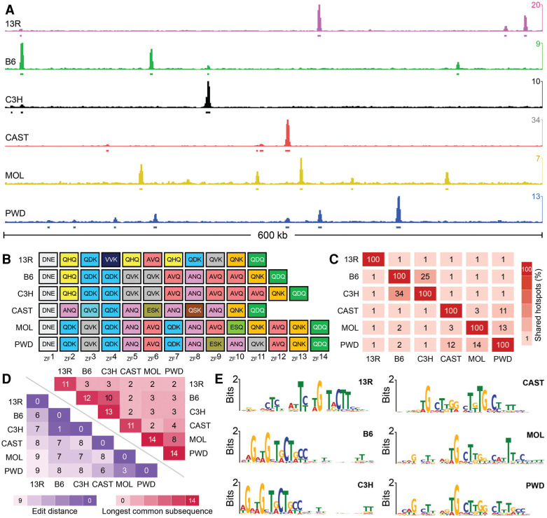
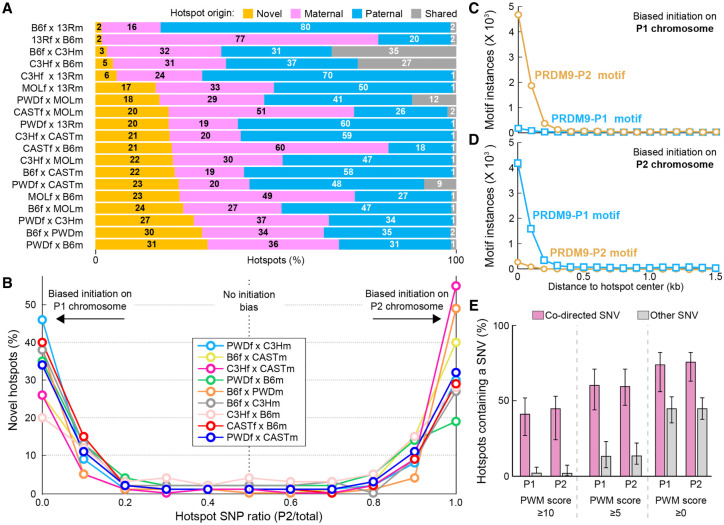
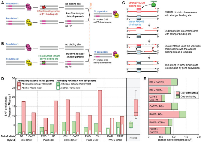
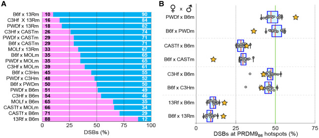
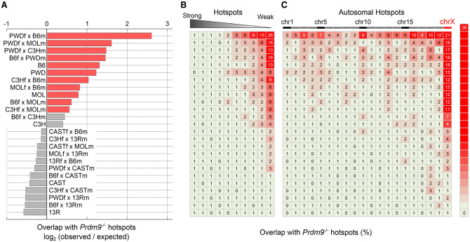
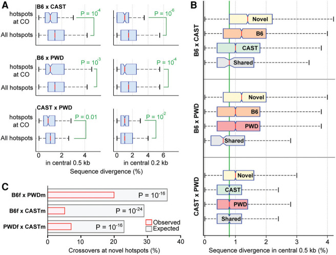
Meiotic recombination is required for the segregation of homologous chromosomes and is essential for fertility. In most mammals, the DNA double-strand breaks (DSBs) that initiate meiotic recombination are directed to a subset of genomic loci (hot spots) by sequence-specific binding of the PRDM9 protein. Rapid evolution of the DNA-binding specificity of PRDM9 and gradual erosion of PRDM9-binding sites by gene conversion will alter the recombination landscape over time. To better understand the evolutionary turnover of recombination hot spots and its consequences, we mapped DSB hot spots in four major subspecies of Mus musculus with different Prdm9 alleles and in their F1 hybrids. We found that hot spot erosion governs the preferential usage of some Prdm9 alleles over others in hybrid mice and increases sequence diversity specifically at hot spots that become active in the hybrids. As crossovers are disfavored at such hot spots, we propose that sequence divergence generated by hot spot turnover may create an impediment for recombination in hybrids, potentially leading to reduced fertility and, eventually, speciation.

Keywords: DSB hot spots; Prdm9; homologous recombination; hybrid sterility; meiosis; recombination hot spots; speciation.


Figures

Figure 1.
Figure 1.
Different alleles of Prdm9 define different DSB hot spots. (A) A snapshot of a 600-kb region on chromosome 1. The Y-axis is given in ssDNA fragments per kilobase per million (FPKM). (B) The ZF array for each Prdm9 allele in this study. ZFs are color-coded by type, and each ZF shows the primary amino acids that confer DNA sequence specificity (positions −1, 3, and 6). (C) The overlap between DSB hot spots in different mouse strains. Overlaps are restricted to the central 400 base pairs (bp) of hot spots. (D) To estimate the similarity of Prdm9 alleles, we considered each allele as a string of independent ZFs and calculated the Damerau-Levenshtein edit distance (blue) and the longest common subsequence shared between alleles (red). The Damerau-Levenshtein edit distance calculates the number of insertions, deletions, and changes required to convert one string to another; thus, lower numbers reflect more similar alleles, as fewer edits are required. A lower edit distance reflects longer shared common subsequences. (E) A single DNA sequence motif is enriched at hot spots defined by each Prdm9 allele.
Figure 2.
Figure 2.
Novel hot spots in F1 hybrid mice. (A) In F1 hybrid mice, up to 31% of hot spots occur at sites that are not used in parental mice (novel hot spots; orange). (B) A majority of novel hot spots exhibit strongly biased DSB formation on one or the other parental chromosome. To generalize findings across multiple strains, we refer to parental chromosomes as P1 (maternal) and P2 (paternal). Initiation biases were determined by examining SNPs between parental genomes. Hot spots were binned in deciles by the fraction of ssDNA-de-rived sequencing reads overlapping SNP loci that contained P2-derived SNPs. (C) P2 PRDM9 motifs are enriched at novel hot spots where DSBs exhibit an initiation bias on the P1 chromosome. (D) P1 PRDM9 motifs are enriched at novel hot spots where DSBs exhibit an initiation bias on the P2 chromosome. (E) A large percentage of hot spots with biased initiation is explained by SNVs between parental genomes. DSB hot spots with an initiation bias were split by initiation bias (P1,P2). The proportion of DSB hot spots that contain a codirected SNV (pink) in the central 500 base pairs (bp) was calculated. Other, noncodirected SNVs were also examined to give an estimate of the expected background variation rate. Bar height represents the average value across all nine F1 strains for which SNV data are available for both parental strains. Error bars represent the maximum and minimum values across all F1 strains. Data for progressively more lenient motif alignment score thresholds are shown from the left to the right panels. The PWM score threshold is surpassed when either parental chromosome harbors a motif that exceeds the score threshold. There is a large excess of codirected SNVs at hot spots that exhibit biased initiation.
Figure 3.
Figure 3.
Sequence variation modulates the DSB hot spot landscape. (A) Appearance of a novel hot spot in the hybrid due to a hot spot-attenuating variant at a PRDM9-binding site in the “self” genome. (B) Appearance of a novel hot spot in the hybrid due to a hot spot-activating variant at a PRDM9-binding site in the “nonself” genome. (C) The mechanism of gene conversion-mediated erosion of PRDM9-binding sites. (D) At novel hot spots, both PRDM9-binding site-activating and -attenuating variants are enriched. We inferred the Prdm9 allele that defined each novel hot spot using the DSB initiation bias. We then inferred the origin of SNPs by comparison across mouse strains (see the Materials and Methods). The SNP density at each hot spot (±250 nt) was compared with that in the flanking region (± 500-nt→2000-nt region), and the enrichment is shown. Solid red bars indicate hot spot-attenuating variants in the “self” lineage. Solid green bars indicate hot spot-activating variants in the “nonself” lineage. Empty bars represent variants assayed at the motif for the other allele of Prdm9 in each hybrid and reflect the variant density at sites not under selection. Both hot spot-activating and -attenuating variants are enriched at novel hot spot centers for all hybrids. (E) We used a motif score threshold of five or greater to assess how many novel hot spots contained only a loss SNP or only a gain SNP in the central 500 bp. SNPs that do not affect motif scores were not considered. On average, loss SNPs are four times more common than gain SNPs.
Figure 4.
Figure 4.
Pseudo-dominance of Prdm9 alleles is dependent on DNA sequence. (A) The proportion of DSBs contributed by each Prdm9 allele was assessed in F1 hybrids. Where possible, novel hot spots were attributed to a parental allele based on the initiation bias at the hot spot. Hot spots that could not be attributed to either allele were not considered (quantified in Supplemental Fig. S12). (B) We quantified the contribution of PRDM9B6 to DSB formation on chromosome X (chrX) in reciprocal crosses (orange stars). The contribution of the B6 allele to DSB formation on the autosomes (gray circles) is also shown as a box plot for each hybrid. In B6×13R and B6×C3H reciprocal crosses, where the two parental X chromosomes are similar, PRDM9B6 contributes equally to DSB formation in both crosses. In hybrids where the parental X chromosomes differ to a greater extent (B6×PWD and B6×CAST), PRDM9B6 contributes fewer DSBs when chrX originated from the B6 strain (B6f). In the case of B6×PWD, this is particularly striking, as PRDM9B6 is pseudo-dominant on chrXPWD (PWDf×B6m) but pseudo-recessive on chrXB6 (B6f×PWDm).
Figure 5.
Figure 5.
Default hot spots are used in wild-type mice. (A) Prdm9-independent default hot spots are used more frequently than expected in 13 strains and hybrids. For each strain/hybrid, the expected overlap was calculated from 1000× randomized sets of hot spots (see the Materials and Methods). Red bars indicate hybrids with significantly more default hot spots than expected (binomial test, Bonferroni cor-rected, P<0.001). Gray bars are not significantly different from expectation. (B) Most default hot spots are weak. DSB hot spots were di-vided into 10 equally sized bins by strength (columns), and the percentage overlap with Prdm9−/− hot spots was calculated for each bin. (C) Default hot spots are particularly prevalent on chrX. The percentage of default hot spots was determined for each chromosome (columns). Default hot spots are also enriched on autosomes in some strains/hybrids. Note that the vertical order of strains and hybrids in A is maintained in B and C.
Figure 6.
Figure 6.
Sequence divergence limits crossover formation. (A) Crossovers (COs) form at less divergent DSB hot spot loci. For this analysis, autosomal DSB hot spots in reciprocal hybrids were merged. Crossover intervals (Liu et al. 2014) that contain a single DSB hot spot were identified, and the diversity at these hot spots was calculated and compared with the diversity at all hot spots. The diversity in different windows around the hot spot center was used (±250 bp in the left panels; ±100 bp in the right panels). Sequence divergence is the percentage of base pairs that differ between parental genomes. Each SNP increased divergence by one, while indels increased divergence by the length of the indel. P-values were calculated using one-tailed Wilcoxon rank-sum test. (B) Divergence between parental genomes is high at novel DSB hot spots. Novel hot spots are significantly more divergent than hot spots defined by either parent in all three hybrids. P<10−4, Wilcoxon rank-sum test. For each hybrid, novel hot spots, hot spots found in each parent, and shared hot spots are shown. The average genome diversity between these strains is 0.8% (green line). (C) Crossovers are significantly depleted at novel hot spots in all three crosses. The expected overlaps were calculated from 10,000× bootstrapped sampling of DSB hot spots. For each iteration, 23 unique F1 DSB hot spots were selected, weighted by hot spot strength. P-values were calculated using a two-sided binomial test.

Similar articles

Cited by

References

    1. Bailey TL, Machanick P. 2012. Inferring direct DNA binding from ChIP-seq. Nucleic Acids Res 40: e128. - PMC - PubMed
    1. Baker CL, Kajita S, Walker M, Saxl RL, Raghupathy N, Choi K, Petkov PM, Paigen K. 2015. PRDM9 drives evolutionary erosion of hotspots in Mus musculus through haplotype-specific initiation of meiotic recombination. PLoS Genet 11: e1004916. - PMC - PubMed
    1. Baudat F, Buard J, Grey C, Fledel-Alon A, Ober C, Przeworski M, Coop G, de Massy B. 2010. PRDM9 is a major determinant of meiotic recombination hotspots in humans and mice. Science 327: 836–840. - PMC - PubMed
    1. Baudat F, Imai Y, de Massy B. 2013. Meiotic recombination in mammals: localization and regulation. Nat Rev Genet 14: 794–806. - PubMed
    1. Berg IL, Neumann R, Lam KW, Sarbajna S, Odenthal-Hesse L, May CA, Jeffreys AJ. 2010. PRDM9 variation strongly influences recombination hot-spot activity and meiotic instability in humans. Nat Genet 42: 859–863. - PMC - PubMed

Publication types

Substances