Review
. 2016 Feb 19;371(1688):20150113.
doi: 10.1098/rstb.2015.0113. Epub 2016 Feb 1.

The importance of having two X chromosomes

Affiliations
Review

The importance of having two X chromosomes

Arthur P Arnold et al. Philos Trans R Soc Lond B Biol Sci. .

Abstract

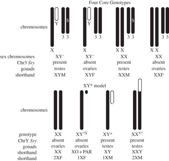
Historically, it was thought that the number of X chromosomes plays little role in causing sex differences in traits. Recently, selected mouse models have been used increasingly to compare mice with the same type of gonad but with one versus two copies of the X chromosome. Study of these models demonstrates that mice with one X chromosome can be strikingly different from those with two X chromosomes, when the differences are not attributable to confounding group differences in gonadal hormones. The number of X chromosomes affects adiposity and metabolic disease, cardiovascular ischaemia/reperfusion injury and behaviour. The effects of X chromosome number are likely the result of inherent differences in expression of X genes that escape inactivation, and are therefore expressed from both X chromosomes in XX mice, resulting in a higher level of expression when two X chromosomes are present. The effects of X chromosome number contribute to sex differences in disease phenotypes, and may explain some features of X chromosome aneuploidies such as in Turner and Klinefelter syndromes.

Keywords: Klinefelter; X chromosome; ischaemia; obesity; sex differences; sexual differentiation.


Figures

Figure 1.
Figure 1.
Effects of one versus two X chromosomes can be revealed using two mouse models that have various combinations of sex chromosomes and gonad type. In the FCG model, the Y chromosome is deleted for Sry, and designated Y. An Sry transgene (S) is present on chromosome 3 in some groups. Breeding XYM with XXF produces the four genotypes, XX and XY mice with testes (XXM, XYM), and XX and XY mice with ovaries (XXF, XYF). In the XY* model, breeding an XX mother with XY* father produces the four genotypes, based on the abnormal recombination of the Y* chromosome with the X chromosome [–45]. Adapted from [27] with permission from Elsevier.
Figure 2.
Figure 2.
The FCG and XY* models are used to demonstrate that the number of X chromosomes contributes to sex differences in body weight and lipoprotein levels. (a) At weaning (age 21 days), the FCG groups have similar body weight, but after puberty (day 45), mice with testes had greater body weight than mice with ovaries (p < 0.000001), and mice with XX sex chromosomes weighed slightly more than XY (*p < 0.05). After mice were gonadectomized (GDX) at 75 days of age, and allowed to grow for 10 months, XX mice weighed much more than XY mice, in both gonadal male groups and gonadal female groups (p < 0.0001), and females were heavier than males (**p < 0.01). The effects of sex chromosome complement interacted significantly with the effects of gonad type (int, *p < 0.05). (b) Growth curves for FCG mice GDX at day 75 (week 0). Before gonadectomy (GDX), mice with ovaries weighed less than mice with testes, and XX mice weighed more than XY mice. The sex difference caused by gonadal secretions acting in adulthood disappeared within four to five weeks after GDX, and thereafter XX mice gained more weight than XY mice. (c) The sex chromosome effect on body weight is confirmed in the XY* model and found to be caused by the number of X chromosomes, using the same GDX design as in a and b. After GDX at day 75, mice with two X chromosomes gained weight more than mice with one X chromosome (p < 0.000001). (d) Metabolic effects of X chromosome number. In FCG mice (left and centre panels) that were gonad-intact or GDX, and in mice eating low-fat lab chow or a high cholesterol diet, XX mice had consistently higher plasma levels of high-density lipoprotein (HDL) cholesterol, independent of their gonadal sex. In the XY* model, mice with two X chromosome had higher HDL levels than mice with one X chromosome. *p < 0.05, **p < 0.01, ***p < 0.001, p < 0.0001. Adapted from [53,54], with permission from Wolters Kluwer Health, Inc.
Figure 3.
Figure 3.
Use of the FCG model shows that after ischaemia/reperfusion injury, GDX XX mice have worse recovery and larger myocardial infarct area compared with GDX XY mice, irrespective of gonadal type. (a) Experimental protocol in vivo: the left anterior descending artery was occluded in GDX FCG mice for 30 min followed by 24 h of reperfusion. (b) Representative cross sections of heart muscle stained with triphenyl tetrazolium chloride. The white area represents the infarcted area, blue shows the non-infarcted area, red plus white areas show risk area. (c) Percentage of area at risk (AAR) divided by left ventricle area. (d) Infarct size (IS) divided by AAR. **p < 0.01, n = 6–7. (e) Experimental protocol ex vivo: perfusion of the heart is shut off for 30 min, then reperfused for 60 min before measuring heart function. (f) The rate pressure product (RPP), a measure of recovery after injury, was worse in XX than XY mice. **p < 0.01. (g) Use of the XY* model shows that in the ex vivo system, mice with two X chromosomes (XX, XXY) have worse recovery (lower RPP) than mice with one X chromosome (XO, XY). (h) The infarct size as the percentage of total ventricular area in hearts ex vivo. *p < 0.05. Adapted from [61] with permission from Oxford University Press.
Figure 4.
Figure 4.
The Sex Chromosome Trisomy model. (a) The model involves crossing FCG XY ¯(Sry+) male (same as XYM in figure 1) with XXY ¯ female who has the same Y ¯ as in the FCG model (figure 1). The father's Sry is transgenic on chromosome 3. Eight genotypes are produced, XX, XY, XXY and XYY, each with either testes or ovaries. (b) Body weight data from SCT mice shows that after gonadectomy (GDX) in adulthood and treatment with testosterone (T), XXY mice weigh more (b) and have more body fat relative to body weight (c), compared with XY mice. Adapted from [77,83].

References

    1. Goodfellow PN, Lovell-Badge R. 1993. SRY and sex determination in mammals. Annu. Rev. Genet. 27, 71–92. (10.1146/annurev.ge.27.120193.000443) - DOI - PubMed
    1. Burgoyne PS. 1998. The role of Y-encoded genes in mammalian spermatogenesis. Semin. Cell Dev. Biol. 9, 423–432. (10.1006/scdb.1998.0228) - DOI - PubMed
    1. Marahrens Y, Panning B, Dausman J, Strauss W, Jaenisch R. 1997. Xist-deficient mice are defective in dosage compensation but not spermatogenesis. Genes Dev. 11, 156–166. (10.1101/gad.11.2.156) - DOI - PubMed
    1. Gupta V, Parisi M, Sturgill D, Nuttall R, Doctolero M, Dudko OK, Malley JD, Eastman PS, Oliver B. 2006. Global analysis of X-chromosome dosage compensation. J. Biol. 5, 3 (10.1186/jbiol30) - DOI - PMC - PubMed
    1. Oliver B. 2007. Sex, dose, and equality. PLoS Biol. 5, e340 (10.1371/journal.pbio.0050340) - DOI - PMC - PubMed

Publication types

Substances