

The effect of low-intensity pulsed ultrasound on bone-tendon junction healing: Initiating after inflammation stage
- PMID: 26833973
- PMCID: PMC6084317
- DOI: 10.1002/jor.23180
The effect of low-intensity pulsed ultrasound on bone-tendon junction healing: Initiating after inflammation stage
Abstract
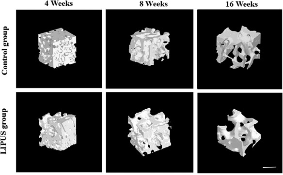
The purpose of this study was to explore the effect of low-intensity pulsed ultrasound (LIPUS) treatment initiating after inflammation stage on the process of bone-tendon junction (BTJ) healing in a rabbit model. Thirty-six rabbits undergoing partial patellectomy were randomly divided into two groups: control and LIPUS. The period of initial inflammatory stage is 2 weeks. So LIPUS treatment was initiated at postoperative week 2 and continued until the patella-patellar tendon (PPT) complexes were harvested at postoperative weeks 4, 8, and 16. At each time point, the PPT complexes were harvested for qRT-PCR, histology, radiographs, synchroton radiation micro computed tomography (SR-µCT), and biomechanical testing. The qRT-PCR results showed that LIPUS treatment beginning at postoperative week 2 played an anti-inflammatory role in BTJ healing. Histologically, the LIPUS group showed more advanced remodeling of the lamellar bone and marrow cavity than the control group. The area and length of the new bone in the LIPUS group were significantly greater than the control group at postoperative weeks 8 and 16. SR-µCT demonstrated that new bone formation and remodeling in the LIPUS group were more advanced than the control group. Biomechanical test results demonstrated that the failure load, ultimate strength and energy at failure were significantly higher than those of the control group. In conclusion, LIPUS treatment beginning at postoperative week 2 was able to accelerate bone formation during the bone-tendon junction healing process and significantly improved the healing quality of BTJ injury. © 2016 Orthopaedic Research Society. Published by Wiley Periodicals, Inc. J Orthop Res 34:1697-1706, 2016.
Keywords: bone-tendon junction (BTJ); low-intensity pulsed ultrasound (LIPUS); synchroton radiation micro computed tomography (SR-µCT).
© 2016 Orthopaedic Research Society. Published by Wiley Periodicals, Inc.
Figures


Similar articles
-
Initiation Timing of Low-Intensity Pulsed Ultrasound Stimulation for Tendon-Bone Healing in a Rabbit Model.Am J Sports Med. 2016 Oct;44(10):2706-2715. doi: 10.1177/0363546516651863. Epub 2016 Jun 29. Am J Sports Med. 2016. PMID: 27358283
-
Combined application of low-intensity pulsed ultrasound and functional electrical stimulation accelerates bone-tendon junction healing in a rabbit model.J Orthop Res. 2014 Feb;32(2):204-9. doi: 10.1002/jor.22505. Epub 2013 Oct 17. J Orthop Res. 2014. PMID: 24136665
-
Effects of low-intensity pulsed ultrasound on new trabecular bone during bone-tendon junction healing in a rabbit model: a synchrotron radiation micro-CT study.PLoS One. 2015 Apr 15;10(4):e0124724. doi: 10.1371/journal.pone.0124724. eCollection 2015. PLoS One. 2015. PMID: 25874957 Free PMC article.
-
The effects of LIPUS on soft-tissue healing: a review of literature.Br Med Bull. 2009;89:169-82. doi: 10.1093/bmb/ldn040. Epub 2008 Nov 16. Br Med Bull. 2009. PMID: 19011263 Review.
-
Low-intensity pulsed ultrasound therapy: a potential strategy to stimulate tendon-bone junction healing.J Zhejiang Univ Sci B. 2012 Dec;13(12):955-63. doi: 10.1631/jzus.B1200129. J Zhejiang Univ Sci B. 2012. PMID: 23225850 Free PMC article. Review.
Cited by
-
SR-FTIR as a tool for quantitative mapping of the content and distribution of extracellular matrix in decellularized book-shape bioscaffolds.BMC Musculoskelet Disord. 2018 Jul 18;19(1):220. doi: 10.1186/s12891-018-2149-9. BMC Musculoskelet Disord. 2018. PMID: 30021603 Free PMC article.
-
Treatment effect of low intensity pulsed ultrasound on leukopenia induced by cyclophosphamide in rabbits.Am J Transl Res. 2017 Jul 15;9(7):3315-3325. eCollection 2017. Am J Transl Res. 2017. PMID: 28804549 Free PMC article.
-
The effects of primary cilia-mediated mechanical stimulation on nestin+-BMSCs during bone-tendon healing.J Adv Res. 2025 Aug;74:415-427. doi: 10.1016/j.jare.2024.09.012. Epub 2024 Sep 19. J Adv Res. 2025. PMID: 39306273 Free PMC article.
-
Strategies for promoting tendon-bone healing: Current status and prospects.Front Bioeng Biotechnol. 2023 Jan 27;11:1118468. doi: 10.3389/fbioe.2023.1118468. eCollection 2023. Front Bioeng Biotechnol. 2023. PMID: 36777256 Free PMC article. Review.
-
Ultrasound as a stimulus for musculoskeletal disorders.J Orthop Translat. 2017 Apr 5;9:52-59. doi: 10.1016/j.jot.2017.03.004. eCollection 2017 Apr. J Orthop Translat. 2017. PMID: 29662799 Free PMC article. Review.
References
-
- Saltzman CL, Goulet JA, McClellan RT, et al. 1990. Results of treatment of displaced patellar fractures by partial patellectomy. J Bone Joint Surg Am 72:1279–1285. - PubMed
-
- Hung LK, Lee SY, Leung KS, et al. 1993. Partial patellectomy for patellar fracture: tension band wiring and early mobilization. J Orthop Trauma 7:252–260. - PubMed
-
- Qin L, Leung KS, Chan CW, et al. 1999. Enlargement of remaining patella after partial patellectomy in rabbits. Med Sci Sports Exerc 31:502–506. - PubMed
-
- Wang L, Qin L, Cheung WH, et al. 2010. A delayed bone‐tendon junction healing model established for potential treatment of related sports injuries. Br J Sports Med 44:114–120. - PubMed
Publication types
MeSH terms
LinkOut - more resources
Full Text Sources
Other Literature Sources
Research Materials
