

Age Differences in Prefrontal Surface Area and Thickness in Middle Aged to Older Adults
- PMID: 26834623
- PMCID: PMC4717301
- DOI: 10.3389/fnagi.2015.00250
Age Differences in Prefrontal Surface Area and Thickness in Middle Aged to Older Adults
Abstract
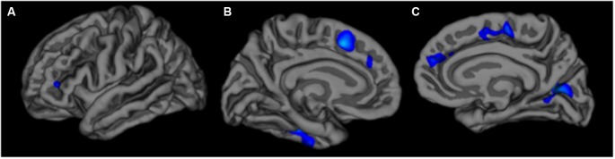
Age is associated with reductions in surface area and cortical thickness, particularly in prefrontal regions. There is also evidence of greater thickness in some regions at older ages. Non-linear age effects in some studies suggest that age may continue to impact brain structure in later decades of life, but relatively few studies have examined the impact of age on brain structure within middle-aged to older adults. We investigated age differences in prefrontal surface area and cortical thickness in healthy adults between the ages of 51 and 81 years. Participants received a structural 3-Tesla magnetic resonance imaging scan. Based on a priori hypotheses, primary analyses focused on surface area and cortical thickness in the dorsolateral prefrontal cortex, anterior cingulate cortex, and orbitofrontal cortex. We also performed exploratory vertex-wise analyses of surface area and cortical thickness across the entire cortex. We found that older age was associated with smaller surface area in the dorsolateral prefrontal and orbitofrontal cortices but greater cortical thickness in the dorsolateral prefrontal and anterior cingulate cortices. Vertex-wise analyses revealed smaller surface area in primarily frontal regions at older ages, but no age effects were found for cortical thickness. Results suggest age is associated with reduced surface area but greater cortical thickness in prefrontal regions during later decades of life, and highlight the differential effects age has on regional surface area and cortical thickness.
Keywords: MRI; adult age differences; aging; cortical thinning; volumetrics.
Figures

Similar articles
-
The structure of the cerebral cortex across adult life: age-related patterns of surface area, thickness, and gyrification.Cereb Cortex. 2013 Nov;23(11):2521-30. doi: 10.1093/cercor/bhs231. Epub 2012 Aug 14. Cereb Cortex. 2013. PMID: 22892423
-
Depressive symptom severity is associated with increased cortical thickness in older adults.Int J Geriatr Psychiatry. 2016 Apr;31(4):325-33. doi: 10.1002/gps.4324. Epub 2015 Jul 23. Int J Geriatr Psychiatry. 2016. PMID: 26205176 Free PMC article.
-
Differential longitudinal changes in cortical thickness, surface area and volume across the adult life span: regions of accelerating and decelerating change.J Neurosci. 2014 Jun 18;34(25):8488-98. doi: 10.1523/JNEUROSCI.0391-14.2014. J Neurosci. 2014. PMID: 24948804 Free PMC article.
-
Bipolar II disorder is associated with thinning of prefrontal and temporal cortices involved in affect regulation.Bipolar Disord. 2013 Dec;15(8):855-64. doi: 10.1111/bdi.12117. Epub 2013 Aug 27. Bipolar Disord. 2013. PMID: 23980618
-
Regionally localized thinning of the cerebral cortex in schizophrenia.Arch Gen Psychiatry. 2003 Sep;60(9):878-88. doi: 10.1001/archpsyc.60.9.878. Arch Gen Psychiatry. 2003. PMID: 12963669
Cited by
-
Brain Shape Changes Associated With Cerebral Atrophy in Healthy Aging and Alzheimer's Disease.Front Mech Eng. 2021 Jul;7:705653. doi: 10.3389/fmech.2021.705653. Epub 2021 Jul 19. Front Mech Eng. 2021. PMID: 35465618 Free PMC article.
-
Cortical aging - new insights with multiparametric quantitative MRI.Aging (Albany NY). 2020 Aug 27;12(16):16195-16210. doi: 10.18632/aging.103629. Epub 2020 Aug 27. Aging (Albany NY). 2020. PMID: 32852283 Free PMC article.
-
Decoupling of mRNA and Protein Expression in Aging Brains Reveals the Age-Dependent Adaptation of Specific Gene Subsets.Cells. 2023 Feb 14;12(4):615. doi: 10.3390/cells12040615. Cells. 2023. PMID: 36831282 Free PMC article.
-
Dynamic changes of region-specific cortical features and scalp-to-cortex distance: implications for transcranial current stimulation modeling.J Neuroeng Rehabil. 2021 Jan 4;18(1):2. doi: 10.1186/s12984-020-00764-5. J Neuroeng Rehabil. 2021. PMID: 33397402 Free PMC article.
-
Effect of dorsolateral prefrontal cortex structural measures on neuroplasticity and response to paired-associative stimulation in Alzheimer's dementia.Philos Trans R Soc Lond B Biol Sci. 2024 Jul 29;379(1906):20230233. doi: 10.1098/rstb.2023.0233. Epub 2024 Jun 10. Philos Trans R Soc Lond B Biol Sci. 2024. PMID: 38853564 Free PMC article.
References
-
- Brandt J., Spencer M., Folstein M. (1988). The telephone interview for cognitive status. Neuropsychiatry Neuropsychol. Behav. Neurol. 1, 111–117.
Grants and funding
LinkOut - more resources
Full Text Sources
Other Literature Sources