Review
. 2016 Jan 18:6:659.
doi: 10.3389/fimmu.2015.00659. eCollection 2015.

Evasion of the Immune Response by Trypanosoma cruzi during Acute Infection

Affiliations
Review

Evasion of the Immune Response by Trypanosoma cruzi during Acute Infection

Mariana S Cardoso et al. Front Immunol. .

Abstract

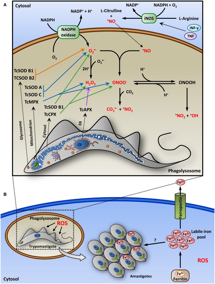
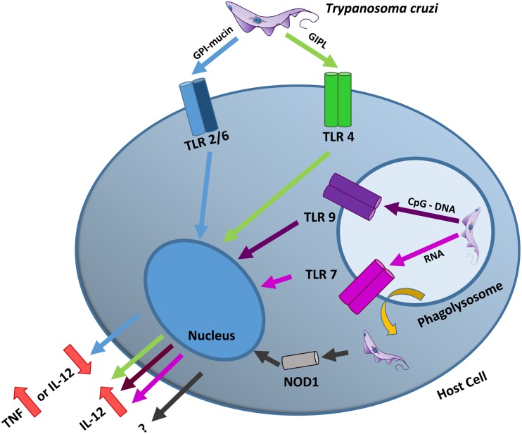
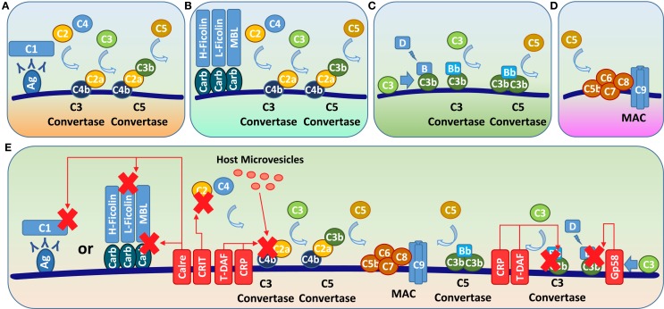
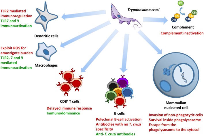
Trypanosoma cruzi is the etiologic agent of Chagas disease, a neglected tropical disease that affects millions of people mainly in Latin America. To establish a life-long infection, T. cruzi must subvert the vertebrate host's immune system, using strategies that can be traced to the parasite's life cycle. Once inside the vertebrate host, metacyclic trypomastigotes rapidly invade a wide variety of nucleated host cells in a membrane-bound compartment known as the parasitophorous vacuole, which fuses to lysosomes, originating the phagolysosome. In this compartment, the parasite relies on a complex network of antioxidant enzymes to shield itself from lysosomal oxygen and nitrogen reactive species. Lysosomal acidification of the parasitophorous vacuole is an important factor that allows trypomastigote escape from the extremely oxidative environment of the phagolysosome to the cytoplasm, where it differentiates into amastigote forms. In the cytosol of infected macrophages, oxidative stress instead of being detrimental to the parasite, favors amastigote burden, which then differentiates into bloodstream trypomastigotes. Trypomastigotes released in the bloodstream upon the rupture of the host cell membrane express surface molecules, such as calreticulin and GP160 proteins, which disrupt initial and key components of the complement pathway, while others such as glycosylphosphatidylinositol-mucins stimulate immunoregulatory receptors, delaying the progression of a protective immune response. After an immunologically silent entry at the early phase of infection, T. cruzi elicits polyclonal B cell activation, hypergammaglobulinemia, and unspecific anti-T. cruzi antibodies, which are inefficient in controlling the infection. Additionally, the coexpression of several related, but not identical, epitopes derived from trypomastigote surface proteins delays the generation of T. cruzi-specific neutralizing antibodies. Later in the infection, the establishment of an anti-T. cruzi CD8(+) immune response focused on the parasite's immunodominant epitopes controls parasitemia and tissue infection, but fails to completely eliminate the parasite. This outcome is not detrimental to the parasite, as it reduces host mortality and maintains the parasite infectivity toward the insect vectors.

Keywords: Chagas disease; T. cruzi acute infection; T. cruzi immune evasion; immune response; immunomodulation.


Figures

Figure 1
Figure 1
Role of host-derived nitroxidative stress in T. cruzi infection. (A) After the phagocytosis of the parasite, macrophage membrane-associated NADPH oxidase is activated, producing the superoxide radical (O2) that can be converted into H2O2 inside the lumen of the phagolysosome. Macrophages stimulated with proinflammatory cytokines (IFN-γ and TNF) induce the expression of nitric oxide synthase (iNOS), generating nitric oxide (NO) in the cytoplasm from the oxidation of l-arginine. NO then diffuse into the phagolysosome vacuole and react with O2 to form peroxynitrite (ONOO), a potent oxidant. Secondary free radicals, such as carbonate (CO3), nitrogen dioxide (NO2), and hydroxyl (OH) radicals, are produced from ONOO. These reactive oxygen species (ROS, indicated in red) can cause various cellular damages and parasite death within the phagolysosome. To survive in this highly oxidative environment, the parasite has a complex network of antioxidant enzymes, as peroxidases (TcAPX, TcCPX, and TcMPX) and superoxide dismutases (SOD), which act in the detoxification of ROS, and are distributed in various cellular compartments, such as glycosomes, mitochondrion, cytosol, and endoplasmic reticulum (ER). Enzymes derived from glycosome, mitochondrion, cytosol, and ER are indicated by orange, blue, green, and purple arrows, respectively. (B) To establish a productive infection, trypomastigotes should escape from the phagolysosome to the cytosol, where it differentiates into replicative amastigotes. In the cytosol of macrophages, ROS, instead of being detrimental to the parasite, can promote the intracellular growth of T. cruzi by a mechanism that may involve facilitating amastigote access to iron. In the cytosol, iron can be stored as ferric iron (Fe3+), a redox-inert form, associated with ferritin or can be exported from the cell as ferrous iron (Fe2+) through ferroportin, a macrophage-specific iron exporter. The expression of ferroportin and ferritin is upregulated by antioxidants, which can lead to reduced levels of labile iron pool in the cytosol. The mechanism of iron uptake by amastigotes is unknown, but the parasite may be dependent on the intracellular labile iron pool for growth.
Figure 2
Figure 2
T. cruzi TLR and NLR activation. T. cruzi possesses several molecules capable of stimulating TLRs. The activation of the heterodimer TLR2/6 by parasite GPI-mucins can lead to TNF production in macrophages or to the inhibition of IL-12 in dendritic cells (blue arrows). By contrast, the activation of TLR4 by parasite GIPLs (green arrows), TLR9 by parasite CpG DNA motifs (purple arrow) and TLR7 by parasite RNA (pink arrow) all result in the production of proinflammatory cytokines, such as IL-12. After the parasite escapes from the phagolysosome, it can activate the cytoplasmic NOD1 receptor. Although this receptor is important for controlling the infection, its mechanism of action is still unknown.
Figure 3
Figure 3
T. cruzi complement evasion mechanisms. There are three complement pathways: classic, alternative, and lectin. (A) In the classical pathway, antibodies bound to pathogen antigens interact with the complement C1 protein, which cleaves C2 and C4 to generate C2a and C4b; these molecules bind to the pathogen surface to form the C3 convertase C4b2a. (B) In the lectin pathway, MBL or ficolin binds to mannan or glycosylated molecules, respectively, on the pathogen surface, and cysteine proteases bound to these molecules cleave C2 and C4, also generating the C3 convertase C4b2a. (C) In the alternative pathway, spontaneously hydrolyzed C3b or C3b originating from the other complement pathways interacts with factor B, which is cleaved into Bb by factor D, forming the C3 convertase C3bBb. The C3 convertases from all the complement pathways interact with newly cleaved C3b, forming a C5 convertase that cleaves C5 into C5b. (D) C5b interacts with C6–C9 to form the MAC, leading to the pathogen lysis. (E) To avoid lysis, T. cruzi relies on molecules, such as calreticulin and gp58/68 (Gp58), which block the initial steps of classic/lectin or alternative pathways, respectively, and CRIT, T-DAF, CRP, and host-derived microvesicles that disrupt or block C3 convertase assembly. Ag, antigen; Carb, carbohydrate; Calre, calreticulin.
Figure 4
Figure 4
Major mechanisms involved in T. cruzi survival and control during the initial phase of infection. This figure summarizes the major interaction mechanisms between T. cruzi and several host components addressed in this review. Mechanisms associated with the control of parasite load are highlighted in green, whereas those involved with parasite modulation of the host immune system and/or with increased parasite load are highlighted in red.

Similar articles

Cited by

References

    1. Hotez PJ, Bottazzi ME, Franco-Paredes C, Ault SK, Periago MR. The neglected tropical diseases of Latin America and the Caribbean: a review of disease burden and distribution and a roadmap for control and elimination. PLoS Negl Trop Dis (2008) 2(9):e300.10.1371/journal.pntd.0000300 - DOI - PMC - PubMed
    1. Coura JR, Dias JC. Epidemiology, control and surveillance of Chagas disease: 100 years after its discovery. Mem Inst Oswaldo Cruz (2009) 104(Suppl 1):31–40.10.1590/S0074-02762009000900006 - DOI - PubMed
    1. Martins-Melo FR, Ramos AN, Jr, Alencar CH, Heukelbach J. Mortality due to Chagas disease in Brazil from 1979 to 2009: trends and regional differences. J Infect Dev Ctries (2012) 6(11):817–24.10.3855/jidc.2459 - DOI - PubMed
    1. Brener Z. Biology of Trypanosoma cruzi. Annu Rev Microbiol (1973) 27:347–82.10.1146/annurev.mi.27.100173.002023 - DOI - PubMed
    1. Stevens JR, Noyes HA, Dover GA, Gibson WC. The ancient and divergent origins of the human pathogenic trypanosomes, Trypanosoma brucei and T. cruzi. Parasitology (1999) 118(Pt 1):107–16.10.1017/S0031182098003473 - DOI - PubMed