Plasmacytoma to the Axis Mimicking Metastatic Paraganglioma: Circumferential Reconstruction via Posterior Approach
- PMID: 26834819
- PMCID: PMC4731566
- DOI: 10.14245/kjs.2015.12.4.283
Plasmacytoma to the Axis Mimicking Metastatic Paraganglioma: Circumferential Reconstruction via Posterior Approach
Abstract
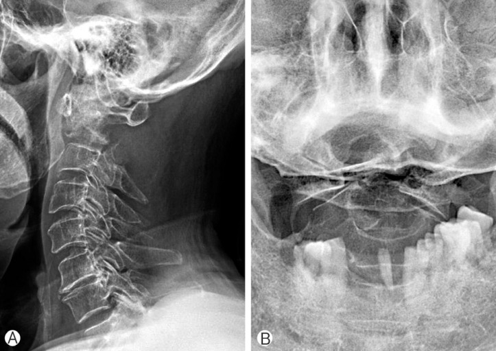
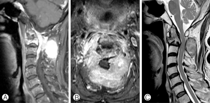
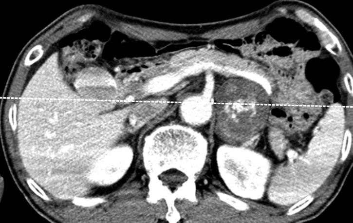
Plasmacytoma is a malignant plasma cell tumor growing within soft tissue or the axial skeleton. Here, we present the case of a patient with plasmacytoma of the axis vertebra who underwent decompressive surgery with reconstruction via a posterior approach. The patient was referred because of quadriparesis with severe neck pain. Magnetic resonance imaging revealed a relatively demarcated, highly enhanced mass lesion in a destructed axis, with spinal cord compression. Computed tomography revealed a 5.6×4.3 cm adrenal mass at the left retroperitoneal space. We suspected the axis lesion to be a metastatic paraganglioma from the adrenal mass. The patient underwent total excision of the tumor under an operative microscope with occipitocervical fixation. Histopathologically, the tumor was shown to be a plasmacytoma. Following the operation, the patient recovered without significant complications. This was a rare case of plasmacytoma in the axis, mimicking metastatic paraganglioma.
Keywords: Axis; Fusion; Paraganglioma; Plasmacytoma; Posterior surgery.
Figures

Similar articles
-
Pediatric cervical kyphosis in the MRI era (1984-2008) with long-term follow up: literature review.Childs Nerv Syst. 2022 Feb;38(2):361-377. doi: 10.1007/s00381-021-05409-z. Epub 2021 Nov 22. Childs Nerv Syst. 2022. PMID: 34806157 Review.
-
Occipitocervical reconstruction through direct lateral and posterior approach for the treatment of primary osteosarcoma in the atlas: a case report.Spine (Phila Pa 1976). 2012 Jan 15;37(2):E126-32. doi: 10.1097/BRS.0b013e31822172b1. Spine (Phila Pa 1976). 2012. PMID: 21629174
-
Paraganglioma presenting as metastatic lesion in a cervical vertebra: a case report and review of the literature.Spine (Phila Pa 1976). 2010 Mar 1;35(5):E152-4. doi: 10.1097/BRS.0b013e3181cf2c96. Spine (Phila Pa 1976). 2010. PMID: 20118832
-
Surgical treatment of malignant paraganglioma with spinal invasion in a juvenile patient: A case report.Medicine (Baltimore). 2019 Sep;98(39):e17145. doi: 10.1097/MD.0000000000017145. Medicine (Baltimore). 2019. PMID: 31574816 Free PMC article.
-
[A case of giant retroperitoneal paraganglioma].Hinyokika Kiyo. 2010 Jul;56(7):377-80. Hinyokika Kiyo. 2010. PMID: 20724811 Review. Japanese.
Cited by
-
Confusion, cognitive impairment, and spinal cord compression caused by plasmacytoma: a case report.BMC Neurol. 2021 Aug 6;21(1):303. doi: 10.1186/s12883-021-02332-3. BMC Neurol. 2021. PMID: 34362322 Free PMC article.
-
Spinal Deformity Surgery : It Becomes an Essential Part of Neurosurgery.J Korean Neurosurg Soc. 2018 Nov;61(6):661-668. doi: 10.3340/jkns.2018.0150. Epub 2018 Oct 30. J Korean Neurosurg Soc. 2018. PMID: 30396240 Free PMC article.
References
-
- Chak LY, Cox RS, Bostwick DG, Hoppe RT. Solitary plasmacytoma of bone: treatment, progression, and survival. J Clin Oncol. 1987;5:1811–1815. - PubMed
-
- Dimopoulos MA, Hamilos G. Solitary bone plasmacytoma and extramedullary plasmacytoma. Curr Treat Options Oncol. 2002;3:255–259. - PubMed
-
- Dimopoulos MA, Moulopoulos LA, Maniatis A, Alexanian R. Solitary plasmacytoma of bone and asymptomatic multiple myeloma. Blood. 2000;96:2037–2044. - PubMed
-
- Grogan TM, Muller-Hermelink HK, Van CB, Harris NL, Kyle RA. Plasm cell neoplasms. In: Jaffe ES, Harris NL, Stein H, Vardiman JW, editors. World health organization classification of tumours. Pathology and genetics of tumours of haematopoietic and lymphoid tissues. Lyon: IARC Press; 2001. pp. 142–156.
-
- Ghogawala Z, Mansfield FL, Borges LF. Spinal radiation before surgical decompression adversely affects outcomes of surgery for symptomatic metastatic spinal cord compression. Spine (Phila Pa 1976) 2001;26:818–824. - PubMed
LinkOut - more resources
Full Text Sources
Other Literature Sources