

Body Surface Electrocardiographic Mapping for Non-invasive Identification of Arrhythmic Sources
- PMID: 26835035
- PMCID: PMC4711575
- DOI: 10.15420/aer.2013.2.1.16
Body Surface Electrocardiographic Mapping for Non-invasive Identification of Arrhythmic Sources
Abstract
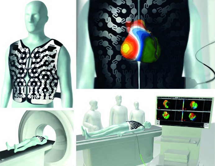
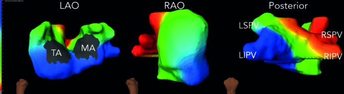
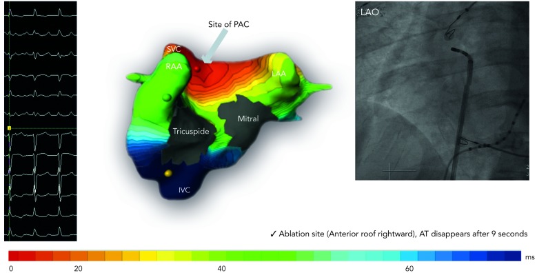
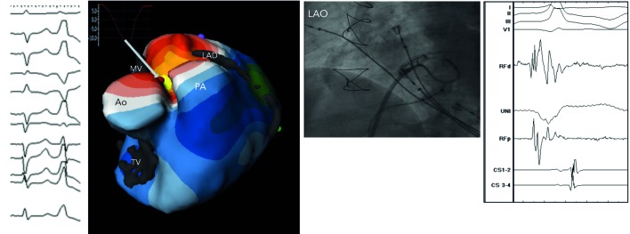
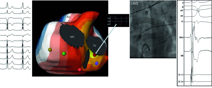
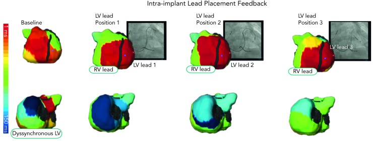
The authors describe a novel three-dimensional, 252-lead electrocardiography (ECG) and computed tomography (CT)-based non-invasive cardiac imaging and mapping modality. This technique images potentials, electrograms and activation sequences (isochrones) on the epicardial surface of the heart. This tool has been investigated in the normal cardiac electrophysiology and various tachyarrhythmic, conduction and anomalous depo-repolarisation disorders. The clinical application of this system includes a wide range of electrical disorders like atrial arrhythmias (premature atrial beat, atrial tachycardia, atrial fibrillation), ventricular arrhythmias (premature ventricular beat, ventricular tachycardia) and ventricular pre-excitation (Wolff-Parkinson-White syndrome). In addition, the system has been used in exploring abnormalities of the His-Purkinje conduction like the bundle branch block and intraventricular conduction disturbance and thereby useful in electrically treating the associated heart failure (cardiac resynchronisation). It has a potential role in furthering our understanding of abnormalities of ventricular action potential (depolarisation [Brugada syndrome and repolarisation], long QT and early repolarisation syndromes) and in evaluating the impact of drugs on His-Purkinje conduction and cardiac action potential.
Keywords: Body surface mapping; arrhythmia sources; electrocardiographic imaging; electrocardiomapping; non-invasive mapping.
Figures

References
-
- The global burden of disease: 2004 update. World Health Organization, Geneva, 2008. Available at: www.who.int/ healthinfo/global_burden_disease/GBD_report_2004update_ ful.... (accessed 25 April 2013).
-
- Oster HS, Taccardi B, Lux RL et al. Noninvasive electrocardiographic imaging: reconstruction of epicardial potentials, electrograms, and isochrones and localization of single and multiple electrocardiac events. Circulation. 1997;96:1012–24. - PubMed
-
- Tikhonov AN, Arsenin VY. Solutions of Ill-Posed Problems. Wiley New York 1977.
-
- Calvetti D, Lewis B, Reichel L. GMRES L-curves and discrete ill-posed problems. BIT. 2002;42:44–65.
-
- Rudy Y, Oster HS. The electrocardiographic inverse problem. Crit Rev Biomed Eng. 1992;20:25–45. - PubMed
LinkOut - more resources
Full Text Sources
Other Literature Sources
