Ovariectomy-Induced Osteoporosis Does Not Impact Fusion Rates in a Recombinant Human Bone Morphogenetic Protein-2-Dependent Rat Posterolateral Arthrodesis Model
- PMID: 26835203
- PMCID: PMC4733376
- DOI: 10.1055/s-0035-1556582
Ovariectomy-Induced Osteoporosis Does Not Impact Fusion Rates in a Recombinant Human Bone Morphogenetic Protein-2-Dependent Rat Posterolateral Arthrodesis Model
Abstract
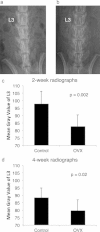
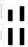
Study Design Randomized, controlled animal study. Objective Recombinant human bone morphogenetic protein-2 (rhBMP-2) is frequently utilized as a bone graft substitute in spinal fusions to overcome the difficult healing environment in patients with osteoporosis. However, the effects of estrogen deficiency and poor bone quality on rhBMP-2 efficacy are unknown. This study sought to determine whether rhBMP-2-induced healing is affected by estrogen deficiency and poor bone quality in a stringent osteoporotic posterolateral spinal fusion model. Methods Aged female Sprague-Dawley rats underwent an ovariectomy (OVX group) or a sham procedure, and the OVX animals were fed a low-calcium, low-phytoestrogen diet. After 12 weeks, the animals underwent a posterolateral spinal fusion with 1 μg rhBMP-2 on an absorbable collagen sponge. Representative animals were sacrificed at 1 week postoperative for alkaline phosphatase (ALP) and osteocalcin serum analyses. The remaining animals underwent radiographs 2 and 4 weeks after surgery and were subsequently euthanized for fusion analysis by manual palpation, micro-computed tomography (CT) imaging, and histologic analysis. Results The ALP and osteocalcin levels were similar between the control and OVX groups. Manual palpation revealed no significant differences in the fusion scores between the control (1.42 ± 0.50) and OVX groups (1.83 ± 0.36; p = 0.07). Fusion rates were 100% in both groups. Micro-CT imaging revealed no significant difference in the quantity of new bone formation, and histologic analysis demonstrated bridging bone across the transverse processes in fused animals from both groups. Conclusions This study demonstrates that estrogen deficiency and compromised bone quality do not negatively influence spinal fusion when utilizing rhBMP-2, and the osteoinductive capacity of the growth factor is not functionally reduced under osteoporotic conditions in the rat. Although osteoporosis is a risk factor for pseudarthrosis/nonunion, rhBMP-2-induced healing was not inhibited in osteoporotic rats.
Keywords: osteoporosis; pseudarthrosis; rats; rhBMP-2; spinal fusion.
Conflict of interest statement
Figures


Similar articles
-
The effect of vancomycin powder on bone healing in a rat spinal rhBMP-2 model.J Neurosurg Spine. 2016 Aug;25(2):147-53. doi: 10.3171/2015.11.SPINE15536. Epub 2016 Apr 1. J Neurosurg Spine. 2016. PMID: 27035510
-
BMP-2 induced early bone formation in spine fusion using rat ovariectomy osteoporosis model.Spine J. 2013 Oct;13(10):1273-80. doi: 10.1016/j.spinee.2013.06.010. Epub 2013 Aug 13. Spine J. 2013. PMID: 23953506
-
The local cytokine and growth factor response to recombinant human bone morphogenetic protein-2 (rhBMP-2) after spinal fusion.Spine J. 2018 Aug;18(8):1424-1433. doi: 10.1016/j.spinee.2018.03.006. Epub 2018 Mar 14. Spine J. 2018. PMID: 29550606
-
rhBMP-2 enhancement of posterolateral spinal fusion in a rabbit model in the presence of concurrently administered doxorubicin.Spine J. 2007 May-Jun;7(3):326-31. doi: 10.1016/j.spinee.2006.06.397. Epub 2006 Dec 22. Spine J. 2007. PMID: 17482116
-
Bone Substitute Options for Spine Fusion in Patients With Spine Trauma-Part II: The Role of rhBMP.Korean J Neurotrauma. 2024 Mar 21;20(1):35-44. doi: 10.13004/kjnt.2024.20.e13. eCollection 2024 Mar. Korean J Neurotrauma. 2024. PMID: 38576507 Free PMC article. Review.
Cited by
-
In situ gel-forming system for dual BMP-2 and 17β-estradiol controlled release for bone regeneration in osteoporotic rats.Drug Deliv Transl Res. 2018 Oct;8(5):1103-1113. doi: 10.1007/s13346-018-0574-9. Drug Deliv Transl Res. 2018. PMID: 30105738
-
Engineered collagen-binding bone morphogenetic protein-2 incorporated with platelet-rich plasma accelerates lumbar fusion in aged rats with osteopenia.Exp Biol Med (Maywood). 2021 Jul;246(14):1577-1585. doi: 10.1177/15353702211001039. Epub 2021 Mar 23. Exp Biol Med (Maywood). 2021. PMID: 33757339 Free PMC article.
-
Optimization of the Time Window of Interest in Ovariectomized Imprinting Control Region Mice for Antiosteoporosis Research.Biomed Res Int. 2017;2017:8417814. doi: 10.1155/2017/8417814. Epub 2017 Oct 8. Biomed Res Int. 2017. PMID: 29119115 Free PMC article.
References
-
- Nelson H D, Haney E M, Dana T, Bougatsos C, Chou R. Screening for osteoporosis: an update for the U.S. Preventive Services Task Force. Ann Intern Med. 2010;153(2):99–111. - PubMed
-
- Burge R, Dawson-Hughes B, Solomon D H, Wong J B, King A, Tosteson A. Incidence and economic burden of osteoporosis-related fractures in the United States, 2005-2025. J Bone Miner Res. 2007;22(3):465–475. - PubMed
-
- HCUP Databases Healthcare Cost and Utilization Project Rockville, MD: Agency for Healthcare Research and Quality. Available at: http://www.hcup-us.ahrq.gov/databases.jsp. Accessed June 21, 2014
-
- Ong K L, Villarraga M L, Lau E, Carreon L Y, Kurtz S M, Glassman S D. Off-label use of bone morphogenetic proteins in the United States using administrative data. Spine (Phila Pa 1976) 2010;35(19):1794–1800. - PubMed
-
- Raizman N M, O'Brien J R, Poehling-Monaghan K L, Yu W D. Pseudarthrosis of the spine. J Am Acad Orthop Surg. 2009;17(8):494–503. - PubMed
LinkOut - more resources
Full Text Sources
Other Literature Sources

