Posterior-Only Circumferential Decompression and Reconstruction in the Surgical Management of Lumbar Vertebral Osteomyelitis
- PMID: 26835214
- PMCID: PMC4733378
- DOI: 10.1055/s-0035-1550341
Posterior-Only Circumferential Decompression and Reconstruction in the Surgical Management of Lumbar Vertebral Osteomyelitis
Abstract
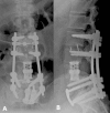
Study Design Case report. Objective The purpose of this report is to discuss the surgical management of lumbar vertebral osteomyelitis with a spinal epidural abscess (SEA) and present a single-stage, posterior-only circumferential decompression and reconstruction with instrumentation using an expandable titanium cage and without segmental nerve root sacrifice as an option in the treatment of this disease process. Methods We report a 42-year-old man who presented with 3 days of low back pain and chills who rapidly decompensated with severe sepsis following admission. Magnetic resonance imaging of his lumbosacral spine revealed intramuscular abscesses of the left paraspinal musculature and iliopsoas with SEA and L4 vertebral body involvement. The patient failed maximal medical treatment, which necessitated surgical treatment as a last resort for infectious source control. He underwent a previously undescribed procedure in the setting of SEA: a single-stage, posterior-only approach for circumferential decompression and reconstruction of the L4 vertebral body with posterior segmental instrumented fixation. Results After the surgery, the patient's condition gradually improved; however, he suffered a wound dehiscence necessitating a surgical exploration and deep wound debridement. Six months after the surgery, the patient underwent a revision surgery for adjacent-level pseudarthrosis. At 1-year follow-up, the patient was pain-free and off narcotic pain medication and had returned to full activity. Conclusion This patient is the first reported case of lumbar osteomyelitis with SEA treated surgically with a single-stage, posterior-only circumferential decompression and reconstruction with posterior instrumentation. Although this approach is more technically challenging, it presents another viable option for the treatment of lumbar vertebral osteomyelitis that may reduce the morbidity associated with an anterior approach.
Keywords: circumferential decompression and reconstruction; lumbar vertebral osteomyelitis; posterior-only approach; spinal epidural abscess.
Conflict of interest statement
Figures


Similar articles
-
The use of an expandable cage for corpectomy reconstruction of vertebral body tumors through a posterior extracavitary approach: a multicenter consecutive case series of prospectively followed patients.Spine J. 2008 Mar-Apr;8(2):329-39. doi: 10.1016/j.spinee.2007.05.002. Epub 2007 Jun 21. Spine J. 2008. PMID: 17923442
-
Single-stage treatment of pyogenic spinal infection with titanium mesh cages.J Spinal Disord Tech. 2006 Jul;19(5):376-82. doi: 10.1097/01.bsd.0000203945.03922.f6. J Spinal Disord Tech. 2006. PMID: 16826013
-
Debridement and Interbody Graft Using Titanium Mesh Cage, Posterior Monosegmental Instrumentation, and Fusion in the Surgical Treatment of Monosegmental Lumbar or Lumbosacral Pyogenic Vertebral Osteomyelitis via a Posterior-Only Approach.World Neurosurg. 2020 Mar;135:e116-e125. doi: 10.1016/j.wneu.2019.11.072. Epub 2019 Nov 20. World Neurosurg. 2020. PMID: 31756509
-
Posterior Nerve-Sparing Multilevel Cervical Corpectomy and Reconstruction for Metastatic Cervical Spine Tumors: Case Report and Literature Review.World Neurosurg. 2019 Feb;122:298-302. doi: 10.1016/j.wneu.2018.11.010. Epub 2018 Nov 14. World Neurosurg. 2019. PMID: 30447451 Review.
-
Spinal epidural abscesses: risk factors, medical versus surgical management, a retrospective review of 128 cases.Spine J. 2014 Feb 1;14(2):326-30. doi: 10.1016/j.spinee.2013.10.046. Epub 2013 Nov 12. Spine J. 2014. PMID: 24231778 Review.
Cited by
-
Clinical results of a lamina with spinous process and an iliac graft as bone grafts in the surgical treatment of single-segment lumbar pyogenic spondylodiscitis: a retrospective cohort study.BMC Surg. 2022 Feb 11;22(1):45. doi: 10.1186/s12893-022-01506-1. BMC Surg. 2022. PMID: 35148743 Free PMC article.
-
Total en bloc spondylectomy in the treatment of postoperative chronic osteomyelitis: a case report.J Spine Surg. 2022 Jun;8(2):288-295. doi: 10.21037/jss-22-14. J Spine Surg. 2022. PMID: 35875627 Free PMC article.
-
One-stage Debridement via Oblique Lateral Interbody Fusion Corridor Combined with Posterior Pedicle Screw Fixation in Treating Spontaneous Lumbar Infectious Spondylodiscitis: A Case Series.Orthop Surg. 2019 Dec;11(6):1109-1119. doi: 10.1111/os.12562. Epub 2019 Nov 7. Orthop Surg. 2019. PMID: 31701667 Free PMC article.
References
-
- Reihsaus E Waldbaur H Seeling W Spinal epidural abscess: a meta-analysis of 915 patients Neurosurg Rev 2000234175–204., discussion 205 - PubMed
-
- Pradilla G, Nagahama Y, Spivak A M, Bydon A, Rigamonti D. Spinal epidural abscess: current diagnosis and management. Curr Infect Dis Rep. 2010;12(6):484–491. - PubMed
-
- Darouiche R O. Spinal epidural abscess. N Engl J Med. 2006;355(19):2012–2020. - PubMed
-
- Davis D P, Wold R M, Patel R J. et al.The clinical presentation and impact of diagnostic delays on emergency department patients with spinal epidural abscess. J Emerg Med. 2004;26(3):285–291. - PubMed
-
- Patel A R, Alton T B, Bransford R J, Lee M J, Bellabarba C B, Chapman J R. Spinal epidural abscesses: risk factors, medical versus surgical management, a retrospective review of 128 cases. Spine J. 2014;14(2):326–330. - PubMed
Publication types
LinkOut - more resources
Full Text Sources
Other Literature Sources

