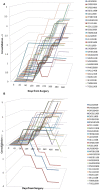
Within Patient Radiological Comparative Analysis of the Performance of Two Bone Graft Extenders Utilized in Posterolateral Lumbar Fusion: A Retrospective Case Series
- PMID: 26835455
- PMCID: PMC4724723
- DOI: 10.3389/fsurg.2015.00069
Within Patient Radiological Comparative Analysis of the Performance of Two Bone Graft Extenders Utilized in Posterolateral Lumbar Fusion: A Retrospective Case Series
Abstract
Two bone graft extenders differing in chemical composition were implanted contralaterally in 27 consecutive patients undergoing instrumented posterolateral lumbar fusion as standard-of-care. Bone marrow aspirate and autogenous bone graft were equally combined either with β-tricalcium phosphate (β-TCP) or a hybrid biomaterial [containing hyaluronic acid (HyA) but lacking a calcium salt] and implanted between the transverse processes. Fusion status on each side of the vertebrae was retrospectively graded (1-5 scale) on AP planar X-ray at multiple visits as available, through approximately 12 months. Additionally, consolidation or resorption since prior visit for each treatment was recorded. Sides receiving β-TCP extender showed marked resorption prior to bone consolidation during the first 6 months. By contrast, sides receiving the hybrid biomaterial containing integrated HyA showed rapid bone consolidation by week 6-8, with maintenance of initial bone volume through 12 months. Fusion grade was superior for the hybrid biomaterial, differing significantly from β-TCP at day 109 and beyond. Fusion success at >12 months was 92.9 vs. 67.9% for the hybrid biomaterial and β-TCP-treated sides, respectively. The hybrid biomaterial extender demonstrated a shortened time-to-fusion compared to the calcium-based graft. Mode of action has been demonstrated in the literature to differ between these compositions. Therefore, choice of synthetic biomaterial composition may significantly influence the mode of action of cellular events regulating appositional bone growth.
Keywords: biomaterial scaffold; bone graft extender; bone marrow aspirate; endochondral ossification; hyaluronic acid; spine fusion.
Figures


Similar articles
-
Influence of 45S5 Bioactive Glass in A Standard Calcium Phosphate Collagen Bone Graft Substitute on the Posterolateral Fusion of Rabbit Spine.Iowa Orthop J. 2017;37:193-198. Iowa Orthop J. 2017. PMID: 28852357 Free PMC article.
-
Single-level instrumented posterolateral fusion of lumbar spine with beta-tricalcium phosphate versus autograft: a prospective, randomized study with 3-year follow-up.Spine (Phila Pa 1976). 2008 May 20;33(12):1299-304. doi: 10.1097/BRS.0b013e3181732a8e. Spine (Phila Pa 1976). 2008. PMID: 18496340 Clinical Trial.
-
A prospective comparative study of radiological outcomes after instrumented posterolateral fusion mass using autologous local bone or a mixture of beta-tcp and autologous local bone in the same patient.Acta Neurochir (Wien). 2013 May;155(5):765-70. doi: 10.1007/s00701-013-1669-1. Epub 2013 Mar 15. Acta Neurochir (Wien). 2013. PMID: 23494134 Clinical Trial.
-
Guideline update for the performance of fusion procedures for degenerative disease of the lumbar spine. Part 16: bone graft extenders and substitutes as an adjunct for lumbar fusion.J Neurosurg Spine. 2014 Jul;21(1):106-32. doi: 10.3171/2014.4.SPINE14325. J Neurosurg Spine. 2014. PMID: 24980593 Review.
-
Experimental posterolateral lumbar spinal fusion with a demineralized bone matrix gel.Spine (Phila Pa 1976). 1998 Jan 15;23(2):159-67. doi: 10.1097/00007632-199801150-00003. Spine (Phila Pa 1976). 1998. PMID: 9474720 Review.
Cited by
-
Synthetic osteobiologics in spine surgery: a review.Turk J Med Sci. 2024 Oct 25;55(1):43-51. doi: 10.55730/1300-0144.5941. eCollection 2025. Turk J Med Sci. 2024. PMID: 40104322 Free PMC article. Review.
-
Use of Autologous Stem Cells in Lumbar Spinal Fusion: A Systematic Review of Current Clinical Evidence.Global Spine J. 2021 Oct;11(8):1281-1298. doi: 10.1177/2192568220973190. Epub 2020 Nov 18. Global Spine J. 2021. PMID: 33203241 Free PMC article.
References
LinkOut - more resources
Full Text Sources
Other Literature Sources

