Case Reports
. 2016 Jan 19:2:73.
doi: 10.3389/fsurg.2015.00073. eCollection 2015.

A Case Report: Pseudoangiomatous Stromal Hyperplasia Tumor Presenting as a Palpable Mass

Affiliations
Case Reports

A Case Report: Pseudoangiomatous Stromal Hyperplasia Tumor Presenting as a Palpable Mass

Q D Vo et al. Front Surg. .

Abstract

We report a case of woman with a palpable lump on her left breast. On mammography, a huge mass located between the inner and the outer inferior breast quadrants of the left breast was found. The ultrasound examination realized later revealed a heterogeneous mass with smooth and lobulated borders. An MRI was also performed, showing an oval mass with heterogeneous areas of enhancement. Finally, a core biopsy under sonographic guidance revealed a pseudoangiomatous stromal hyperplasia of the breast.

Keywords: MRI; breast tumor; mammography; phyllode tumor; pseudoangiomatous stromal hyperplasia.


Figures

Figure 1
Figure 1
Mammography performed with craniocaudal (A) and mediolateral oblique (B) incidences of the left breast shows a huge well-defined mass located between the inner and the outer inferior quadrants, with no suspicious features.
Figure 2
Figure 2
Ultrasound of the left breast centered on the palpable lump showing an oval, heterogeneous lesions with smooth borders.
Figure 3
Figure 3
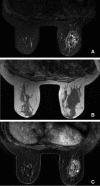
MRI showing the mass on the left breast with lobulated appearances. On T2-weighted MR image with fat saturation (A), the lesion demonstrates a heterogeneous appearance with areas of low and high signal. On T1-weighted MR image (B), the lesion shows a more homogeneous appearance with a hyposignal. On T1-weighted MR image with gadolinium and fat saturation (C), the mass demonstrates a heterogeneous enhancement, with a polylobulated appearance. No lymphadenopathies are seen.
Figure 4
Figure 4
Microscopic examination showing a pseudo vascular proliferation delineated with endothelial cells with canalar hyperplasia without atypia, without invasion of parenchyma.

References

    1. Vuitch MF, Rosen PP, Erlandson RA. Pseudoangiomatous hyperplasia of mammary stroma. Hum Pathol (1986) 17(2):185–91.10.1016/S0046-8177(86)80292-1 - DOI - PubMed
    1. Sasaki Y, Kamata S, Saito K, Nishikawa Y, Ogawa J. Pseudo angiomatous stromal hyperplasia (PASH) of the mammary gland: report of a case. Surg Today (2008) 38(4):340–3.10.1007/s00595-007-3613-6 - DOI - PubMed
    1. Anderson C, Ricci A, Jr, Pedersen CA, Cartun RW. Immunocytochemical analysis of estrogen and progesterone receptors in benign stromal lesions of the breast. Evidence for hormonal etiology in pseudoangiomatous hyperplasia of mammary stroma. Am J Surg Pathol (1991) 15(2):145–9.10.1097/00000478-199102000-00007 - DOI - PubMed
    1. Cohen MA, Morris EA, Rosen PP, Dershaw DD, Liberman L, Abramson AF. Pseudoangiomatous stromal hyperplasia: mammographic, sonographic, and clinical patterns. Radiology (1996) 198(1):117–20. - PubMed
    1. Ferreira M, Albarracin CT, Resetkova E. Pseudoangiomatous stromal hyperplasia tumor: a clinical, radiologic and pathologic study of 26 cases. Mod Pathol (2008) 21(2):201–7.10.1038/modpathol.3801003 - DOI - PubMed

Publication types

LinkOut - more resources