

Quantification of Right and Left Ventricular Function in Cardiac MR Imaging: Comparison of Semiautomatic and Manual Segmentation Algorithms
- PMID: 26835680
- PMCID: PMC4665532
- DOI: 10.3390/diagnostics3020271
Quantification of Right and Left Ventricular Function in Cardiac MR Imaging: Comparison of Semiautomatic and Manual Segmentation Algorithms
Abstract
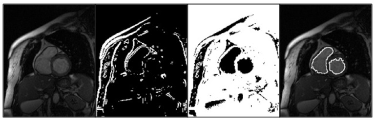
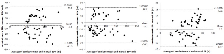
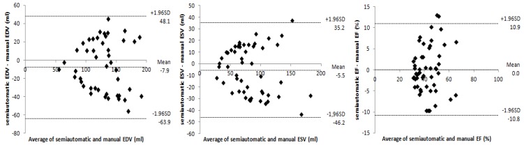
The purpose of this study was to evaluate the performance of a semiautomatic segmentation method for the anatomical and functional assessment of both ventricles from cardiac cine magnetic resonance (MR) examinations, reducing user interaction to a "mouse-click". Fifty-two patients with cardiovascular diseases were examined using a 1.5-T MR imaging unit. Several parameters of both ventricles, such as end-diastolic volume (EDV), end-systolic volume (ESV) and ejection fraction (EF), were quantified by an experienced operator using the conventional method based on manually-defined contours, as the standard of reference; and a novel semiautomatic segmentation method based on edge detection, iterative thresholding and region growing techniques, for evaluation purposes. No statistically significant differences were found between the two measurement values obtained for each parameter (p > 0.05). Correlation to estimate right ventricular function was good (r > 0.8) and turned out to be excellent (r > 0.9) for the left ventricle (LV). Bland-Altman plots revealed acceptable limits of agreement between the two methods (95%). Our study findings indicate that the proposed technique allows a fast and accurate assessment of both ventricles. However, further improvements are needed to equal results achieved for the right ventricle (RV) using the conventional methodology.
Keywords: cardiac cine magnetic resonance imaging (MRI); ejection fraction (EF); left ventricular function; right ventricular function; segmentation.
Figures

References
-
- Becker M., Frauenrath T., Hezel F., Krombach G.A., Kremer U., Koppers B., Butenweg C., Goemmel A., Utting J.F., Schulz-Menger J., Niendorf T. Comparison of left ventricular function assessment using phonocardiogram- and electrocardiogram-triggered 2D SSFP CINE MR imaging at 1.5 T and 3.0 T. Eur. Radiol. 2010;20:1344–1355. doi: 10.1007/s00330-009-1676-z. - DOI - PubMed
-
- Lubbers D.D., Willems T.P., van der Vleuten P.A., Overbosch J., Götte M.J., van Veldhuisen D.J., Oudkerk M. Assessment of global left ventricular functional parameters: Analysis of every second short-axis magnetic resonance imaging slices is as accurate as analysis of consecutive slices. Int. J. Cardiovasc. Imag. 2008;24:185–191. doi: 10.1007/s10554-007-9245-5. - DOI - PubMed
-
- Kunz R.P., Oellig F., Krummenauer F., Oberholzer K., Romaneehsen B., Vomweg T.W., Horstick G., Hayes C., Thelen M., Kreitner K.F. Assessment of left ventricular function by breath-hold cine MR imaging: Comparison of different steady-state free precession sequences. J. Magn. Reson. Imag. 2005;21:140–148. doi: 10.1002/jmri.20230. - DOI - PubMed
LinkOut - more resources
Full Text Sources
Other Literature Sources
