

Evaluation of conventional imaging performance in a research whole-body CT system with a photon-counting detector array
- PMID: 26835839
- PMCID: PMC4782185
- DOI: 10.1088/0031-9155/61/4/1572
Evaluation of conventional imaging performance in a research whole-body CT system with a photon-counting detector array
Abstract
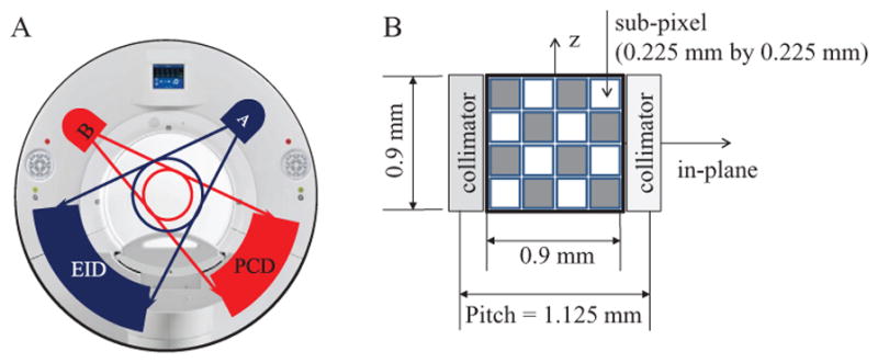
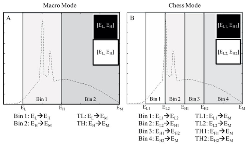
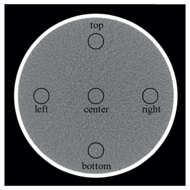
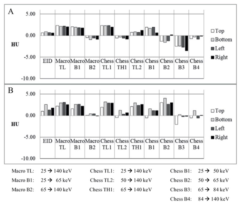
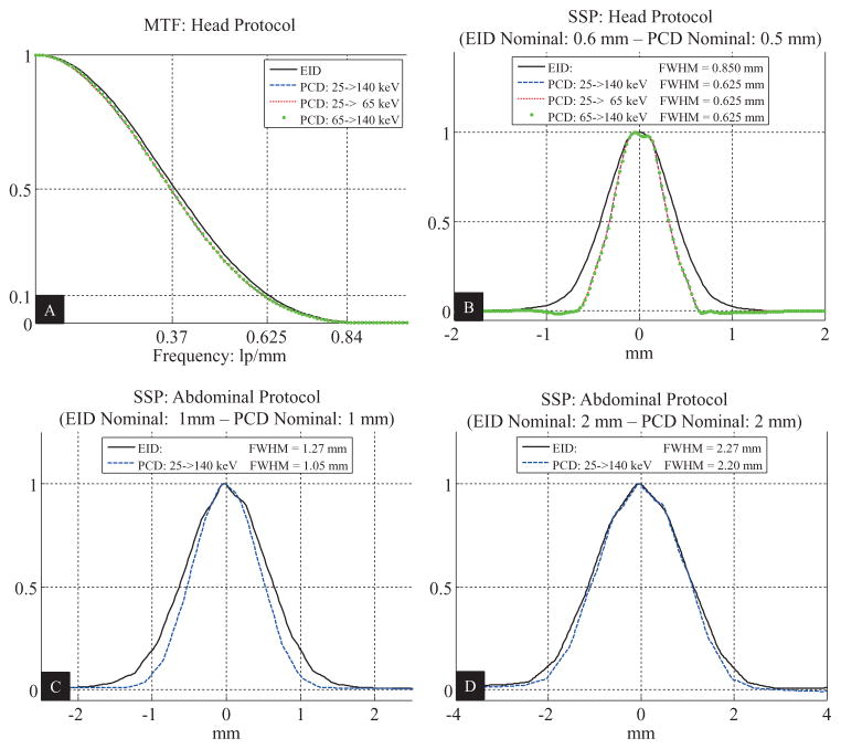
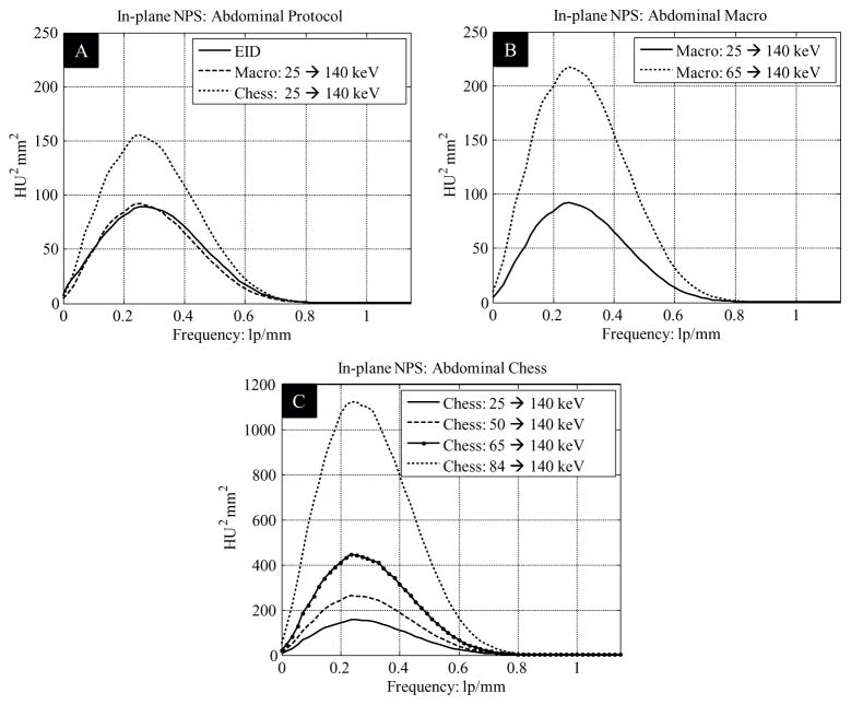
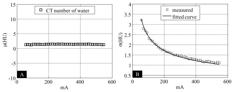
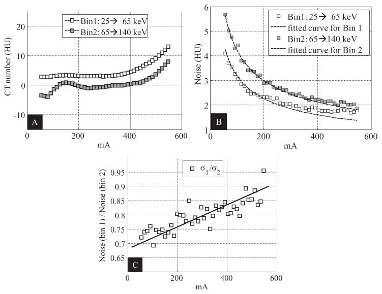
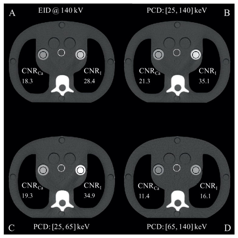
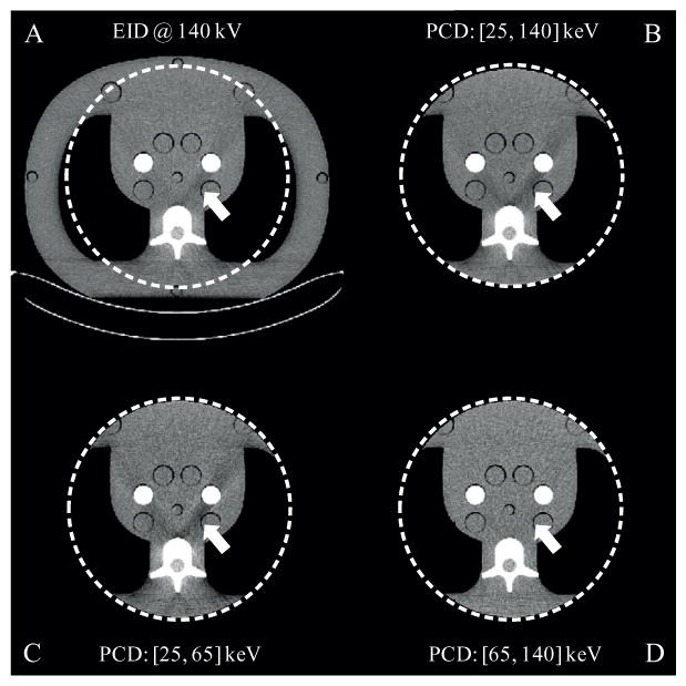
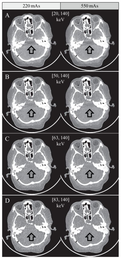
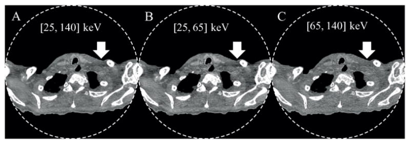
This study evaluated the conventional imaging performance of a research whole-body photon-counting CT system and investigated its feasibility for imaging using clinically realistic levels of x-ray photon flux. This research system was built on the platform of a 2nd generation dual-source CT system: one source coupled to an energy integrating detector (EID) and the other coupled to a photon-counting detector (PCD). Phantom studies were conducted to measure CT number accuracy and uniformity for water, CT number energy dependency for high-Z materials, spatial resolution, noise, and contrast-to-noise ratio. The results from the EID and PCD subsystems were compared. The impact of high photon flux, such as pulse pile-up, was assessed by studying the noise-to-tube-current relationship using a neonate water phantom and high x-ray photon flux. Finally, clinical feasibility of the PCD subsystem was investigated using anthropomorphic phantoms, a cadaveric head, and a whole-body cadaver, which were scanned at dose levels equivalent to or higher than those used clinically. Phantom measurements demonstrated that the PCD subsystem provided comparable image quality to the EID subsystem, except that the PCD subsystem provided slightly better longitudinal spatial resolution and about 25% improvement in contrast-to-noise ratio for iodine. The impact of high photon flux was found to be negligible for the PCD subsystem: only subtle high-flux effects were noticed for tube currents higher than 300 mA in images of the neonate water phantom. Results of the anthropomorphic phantom and cadaver scans demonstrated comparable image quality between the EID and PCD subsystems. There were no noticeable ring, streaking, or cupping/capping artifacts in the PCD images. In addition, the PCD subsystem provided spectral information. Our experiments demonstrated that the research whole-body photon-counting CT system is capable of providing clinical image quality at clinically realistic levels of x-ray photon flux.
Figures
References
-
- Altman A, Carmi R. TU-E-210A-03: a double-layer detector, dual-energy CT—principles, advantages and applications. Med Phys. 2009;36:2750.
-
- Alvarez RE, Macovski A. Energy-selective reconstructions in x-ray computerised tomography. Phys Med Biol. 1976;21:733. - PubMed
-
- Bennett JR, Opie AM, Xu Q, Yu H, Walsh M, Butler A, Butler P, Cao G, Mohs A, Wang G. Hybrid spectral micro-CT: system design, implementation, and preliminary results. IEEE Trans Biomed Eng. 2014;61:246–53. - PubMed
-
- Cody DD, Pfeiffer D, McNitt-Gray MF, Ruckdeschel T, Strauss KJ. CT Quality Control Manual. Reston, VA: American College of Radiology; 2012.
-
- Faby S, Kuchenbecker S, Sawall S, Simons D, Schlemmer H-P, Lell M, Kachelrieß M. Performance of today’s dual energy CT and future multi energy CT in virtual non-contrast imaging and in iodine quantification: a simulation study. Med Phys. 2015;42:4349–66. - PubMed
Publication types
MeSH terms
Grants and funding
LinkOut - more resources
Full Text Sources
Other Literature Sources
Medical