. 2016 Feb;185(2):169-81.
doi: 10.1667/RR14285.1. Epub 2016 Feb 2.

Investigating the Radioresistant Properties of Lung Cancer Stem Cells in the Context of the Tumor Microenvironment

Affiliations

Investigating the Radioresistant Properties of Lung Cancer Stem Cells in the Context of the Tumor Microenvironment

Ryan Chan et al. Radiat Res. 2016 Feb.

Abstract

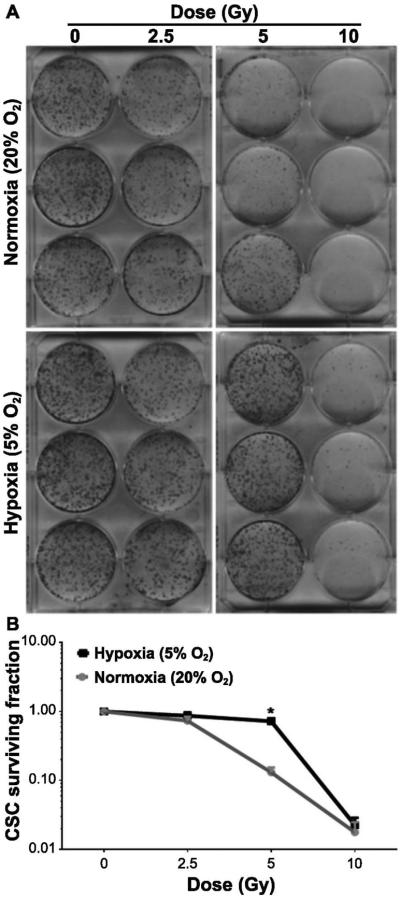
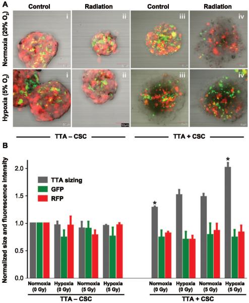
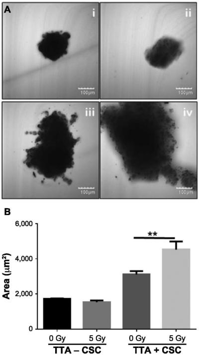
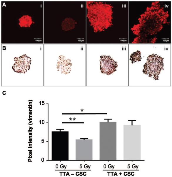
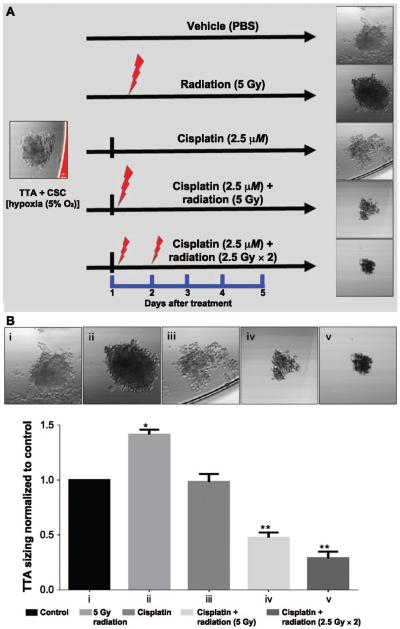
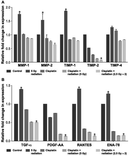
Lung cancer is the most common cause of cancer-related deaths worldwide and non-small cell lung cancer (NSCLC) accounts for ~85% of all lung cancer. While recent research has shown that cancer stem cells (CSC) exhibit radioresistant and chemoresistant properties, current cancer therapy targets the bulk of the tumor burden without accounting for the CSC and the contribution of the tumor microenvironment. CSC interaction with the stroma enhances NSCLC survival, thus limiting the efficacy of treatment. The aim of this study was to elucidate the role of CSC and the microenvironment in conferring radio- or chemoresistance in an in vitro tumor model for NSCLC. The novel in vitro three-dimensional (3D) NSCLC model of color-coded tumor tissue analogs (TTA) that we have developed is comprised of human lung adenocarcinoma cells, fibroblasts, endothelial cells and NSCLC cancer stem cells maintained in low oxygen conditions (5% O2) to recapitulate the physiologic conditions in tumors. Using this model, we demonstrate that a single 5 Gy radiation dose does not inhibit growth of TTA containing CSC and results in elevated expression of cytokines (TGF-α, RANTES, ENA-78) and factors (vimentin, MMP and TIMP), indicative of an invasive and aggressive phenotype. However, combined treatment of single dose or fractionated doses with cisplatin was found to either attenuate or decrease the proliferative effect that radiation exposure alone had on TTA containing CSC maintained in hypoxic conditions. In summary, we utilized a 3D NSCLC model, which had characteristics of the tumor microenvironment and tumor cell heterogeneity, to elucidate the multifactorial nature of radioresistance in tumors.


Figures

FIG. 1
FIG. 1
The hypoxic microenvironment increases CSC radioresistance. Panels A and B: CSC maintained in normoxia (20% O2) and hypoxia (5% O2) were exposed to increasing doses of radiation, as indicated. Cells were further maintained for 14 days after the media was replaced. Colonies (>50 cells) were stained and counted (see Materials and Methods). Data represent means ± SD (n = 3). Significant change in the surviving fraction of the CSC population maintained in hypoxic and normoxic conditions after 5 Gy irradiation (recorded as *P < 0.01).
FIG. 2
FIG. 2
The radiation-induced active proliferation of CSC in tumor tissue analogs (TTA) maintained in hypoxia accounts for the robust growth of TTA. Panel A: Confocal images of TTA maintained in normoxia 20% (upper images) and hypoxia 5% (lower images) with and without radiation treatment. Treatment regimens shown in the upper and lower images, from left to right, are as follows: i. TTA − CSC control; ii. TTA − CSC 5 Gy radiation; iii. TTA + CSC control; and iv. TTA + CSC 5 Gy radiation. Panel B. Graphical representation of size, green and red fluorescence of the TTA normalized to the untreated control (TTA − CSC) maintained in normoxic conditions. While the mere presence of CSC in the TTA under normoxic conditions resulted in a significant increase in size, radiation treatment and hypoxic microenvironment caused a further increase in the size of the TTA + CSC, with *P < 0.005 (n = 3). Abbreviations are as follows: TTA − CSC (A549, endothelial and fibroblast), TTA + CSC (A549, endothelial and fibroblast with CSC). The images are an overlay of red (tumor cells) and green (fibroblasts) fluorescence. The endothelial cells and CSC do not express any fluorescent protein.
FIG. 3
FIG. 3
The presence of CSC in TTA maintained in a hypoxic environment (5% O2) results in increased TTA growth, which is further enhanced after irradiation. Panel A: Phase-contrast images of TTA using confocal microscopy under the following treatment regimens (left to right): i. TTA−CSC control; ii. TTA−CSC 5 Gy radiation; iii. TTA + CSC control; and iv. TTA + CSC 5 Gy radiation. Panel B. Sizing of tumor tissue analogs grown in hypoxia by area analysis conducted in ImageJ. Mean area values are shown. Abbreviations are as follows: TTA−CSC (A549 lung adenocarcinoma cells, pulmonary endothelial cells and fibroblast), TTA + CSC (A549 lung adenocarcinoma cells, pulmonary endothelial and fibroblast with CSC). (n = 4 TTA were utilized per treatment to assess spheroid size). **P < 0.008 indicates a statistically significant difference between the treatments.
FIG. 4
FIG. 4
Radiation treatment did not notably alter the increased expression of vimentin in TTA with CSC grown in hypoxia. TTA − CSC and TTA + CSC maintained in hypoxia at 5 days after 5 Gy irradiation were incubated with rabbit anti-vimentin polyclonal antibody followed by anti-rabbit secondary Alexa Fluor 633 (red, panel A) and monoclonal mouse anti-vimentin (Dako, Inc.) followed by subsequent DAB staining (panel B). Treatment regimens are as follows (left to right for panels A and B): i. TTA − CSC control; ii. TTA − CSC 5 Gy radiation; iii. TTA + CSC control; iv. TTA + CSC 5 Gy radiation. Panel C: Intensity analysis of antibody staining for vimentin in the respective groups shown in panel A. The average pixel intensity for vimentin in the different treatment groups was analyzed using ImageJ software. Mean ratio values are shown. Abbreviations are as follows: −CSC (A549, endothelial, fibroblast), +CSC (A549, endothelial, fibroblast, CSC). (n = 4 TTA were used per treatment assess spheroid size.) **P < 0.009 and *P < 0.01 indicate a statistically significant difference between the treatments.
FIG. 5
FIG. 5
Combined cisplatin and fractionated radiation treatment inhibits the growth of TTA + CSC in a hypoxic environment. Panel A: A schematic representation of TTA + CSC maintained in the hypoxic microenvironment and treated with different combinations of radiation and/or cisplatin. Panel B: Confocal phase-contrast images (upper images) of TTA + CSC grown in hypoxia (5%) after various treatment regimens (left to right): i. control; ii. single 5 Gy dose; iii. 2.5 μM cisplatin; iv. 2.5 μM cisplatin + single 5 Gy dose; v. 2.5 μM cisplatin + fractionated doses (2.5 Gy × 2). The images were taken on treatment day 5. ImageJ software analysis of sizing in TTA + CSC maintained in hypoxia revealed a significant decrease (P < 0.01) with combined cisplatin and single dose or fractionated doses compared to radiation treatment alone.
FIG. 6
FIG. 6
Single doses or fractionated doses combined with cisplatin in the TTA + CSC reverts the expression profile of proteins and cytokines associated with the invasive phenotype in lung cancer. Panel A: Quantification of pixel intensity as a measure of protein expression from TTA + CSC lysates 5 days after treatment incubated with the human MMP and TIMP multiplex antibody arrays. Panel B: Quantification of pixel intensity as a measure of protein expression from the culture media maintaining TTA + CSC 5 days after treatment and incubated with the human cytokine/chemokine antibody arrays. The results are presented as mean ± SE of three different determinations. *Significant difference (P < 0.05) in the treatment group compared to the untreated control group.

References

    1. Cancer Facts and Figures. American Cancer Society; 2015. bit.ly/1tMudoF.
    1. Price A. Emerging developments of chemoradiotherapy in stage III NSCLC. Nat Rev Clin Oncol. 2012;9:591–8. - PubMed
    1. Jett JR, Schild SE, Keith RL, Kesler KA, American College of Chest Physicians Treatment of non-small cell lung cancer, stage IIIB: ACCP evidence-based clinical practice guidelines (2nd edition) Chest. 2007;132:266S–76S. - PubMed
    1. Robinson LA, Ruckdeschel JC, Wagner H, Jr., Stevens CW, American College of Chest Physicians Treatment of non-small cell lung cancer-stage IIIA: ACCP evidence-based clinical practice guidelines (2nd edition) Chest. 2007;132:243S–65S. - PubMed
    1. Auperin A, Le Pechoux C, Rolland E, Curran WJ, Furuse K, Fournel P, et al. Meta-analysis of concomitant versus sequential radiochemotherapy in locally advanced non-small-cell lung cancer. J Clin Oncol. 2010;28:2181–90. - PubMed

Publication types

MeSH terms