

Changes in patellofemoral and tibiofemoral joint cartilage damage and bone marrow lesions over 7 years: the Multicenter Osteoarthritis Study
- PMID: 26836287
- PMCID: PMC4907825
- DOI: 10.1016/j.joca.2016.01.981
Changes in patellofemoral and tibiofemoral joint cartilage damage and bone marrow lesions over 7 years: the Multicenter Osteoarthritis Study
Abstract
Objectives: To investigate changes in cartilage damage and bone marrow lesions (BMLs) on MRI in the patellofemoral and tibiofemoral joints (TFJs) over 7 years.
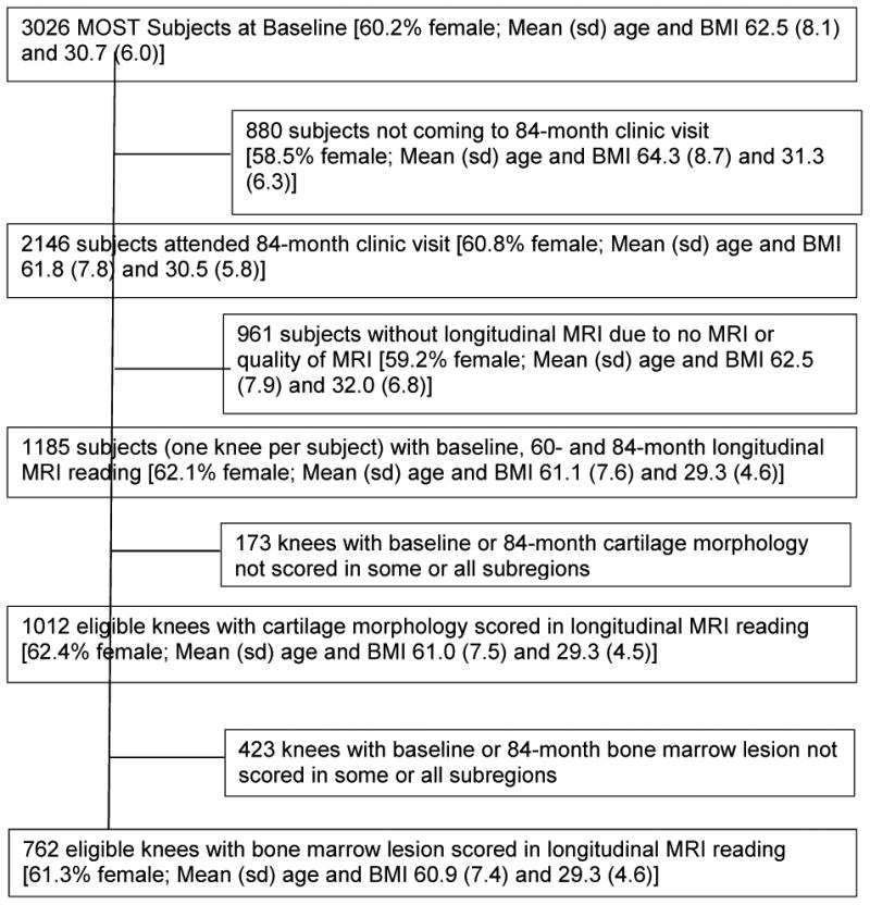
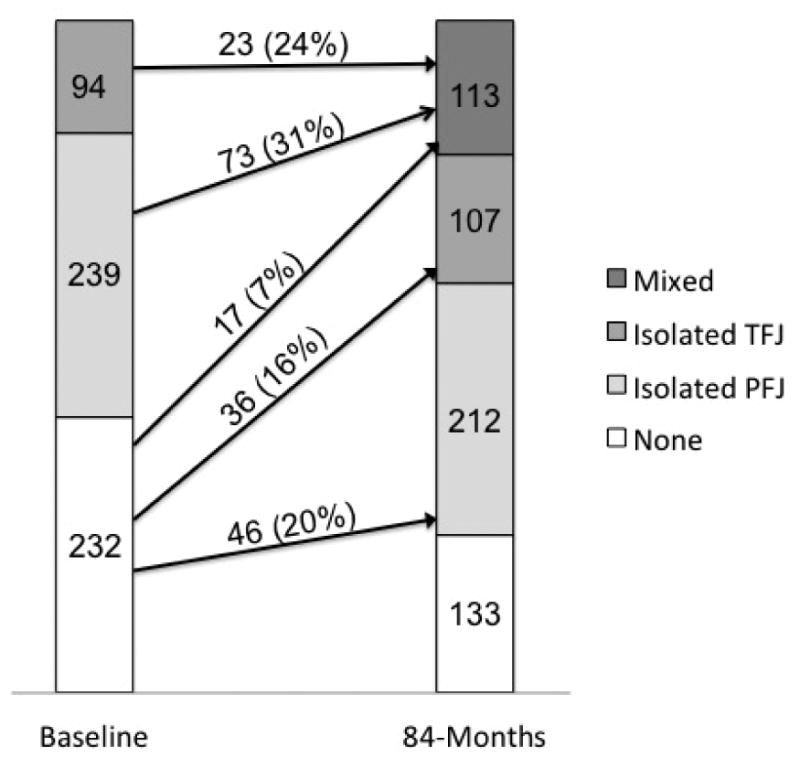
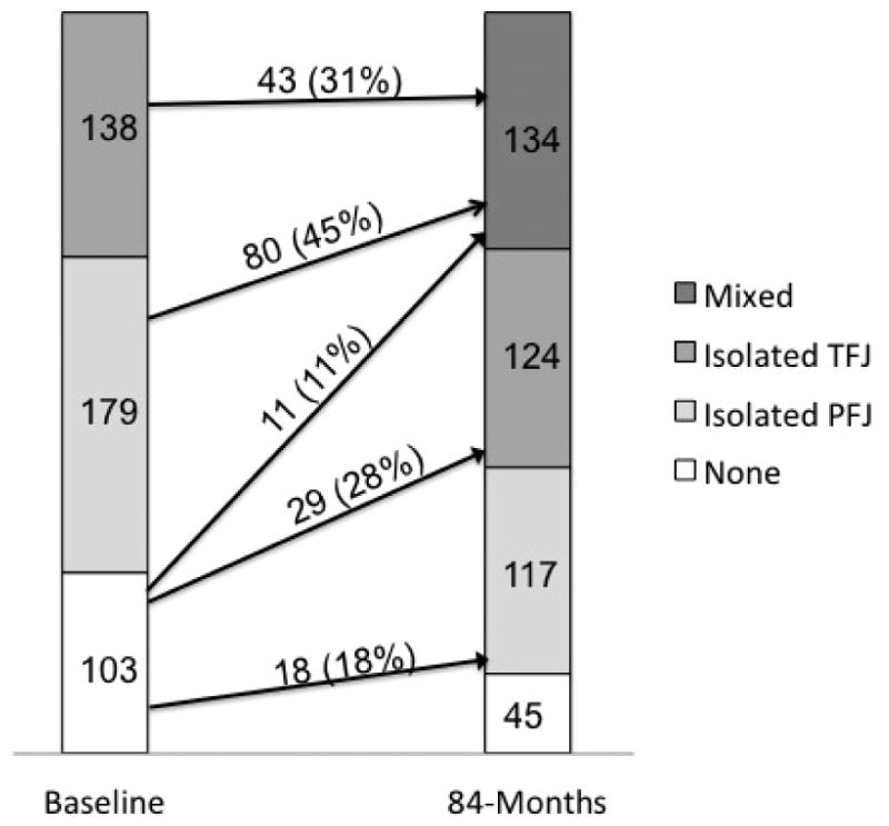
Methods: The Multicenter Osteoarthritis (MOST) Study is a cohort study of persons aged 50-79 years at baseline with or at high risk for knee osteoarthritis (OA). Knees were eligible for the current study if they had knee MRI (1.0T) assessed for cartilage damage and BMLs at the baseline and 84-month visits. Knees were categorized as having MRI-detected structural damage (cartilage and BMLs) isolated to the patellofemoral joint (PFJ), isolated to the TFJ, mixed or no damage at baseline and 84-months. We determined the changes in PFJ and TFJ structural damage over 7 years and used logistic regression to assess the relation of baseline compartment distribution to incident isolated PFJ, isolated TFJ and mixed damage.
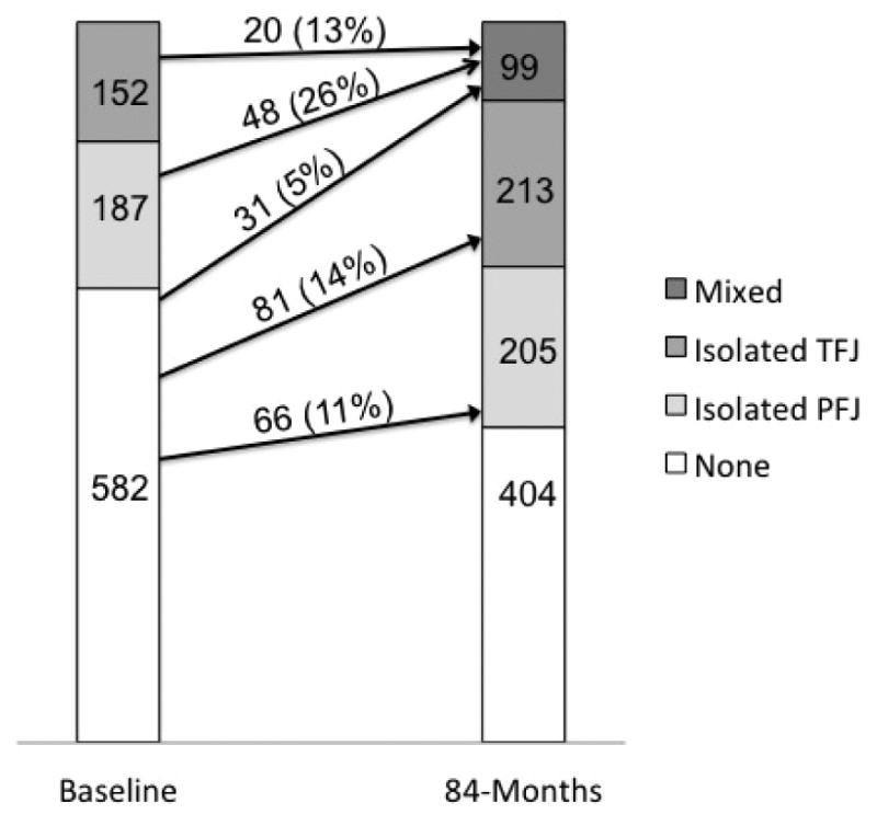
Results: Among 339 knees that had full-thickness cartilage loss isolated to the PFJ or TFJ at baseline, only 68 (20.1%) developed full-thickness cartilage loss in the other compartment while 271 (79.9%) continued to only have the initial compartment affected. Compared to knees without full-thickness cartilage damage (n = 582), those with isolated TFJ and PFJ full-thickness cartilage damage had 2.7 (1.5, 4.9) and 5.8 (3.6, 9.6) times the odds of incident mixed full-thickness cartilage damage, respectively. Similar results were seen when using other definitions of MRI-defined structural damage.
Conclusions: Most knees with structural damage at baseline do not develop it in the other compartment. Knees that develop mixed structural damage are more likely to start with it isolated to the PFJ.
Keywords: Knee osteoarthritis; MRI; Pain.
Copyright © 2016 Osteoarthritis Research Society International. Published by Elsevier Ltd. All rights reserved.
Figures
References
-
- Crossley KM, Marino GP, Macilquham MD, Schache AG, Hinman RS. Can patellar tape reduce the patellar malalignment and pain associated with patellofemoral osteoarthritis? Arthritis Rheum. 2009;61(12):1719–25. - PubMed
-
- Duncan R, Peat G, Thomas E, Hay EM, Croft P. Incidence, progression and sequence of development of radiographic knee osteoarthritis in a symptomatic population. Ann Rheum Dis. 2011;70(11):1944–8. - PubMed
Publication types
MeSH terms
Grants and funding
LinkOut - more resources
Full Text Sources
Other Literature Sources
