. 2016 Jan;8(1):192-207.
doi: 10.18632/aging.100876.

miR-320a mediates doxorubicin-induced cardiotoxicity by targeting VEGF signal pathway

Affiliations

miR-320a mediates doxorubicin-induced cardiotoxicity by targeting VEGF signal pathway

Zhongwei Yin et al. Aging (Albany NY). 2016 Jan.

Erratum in

Abstract

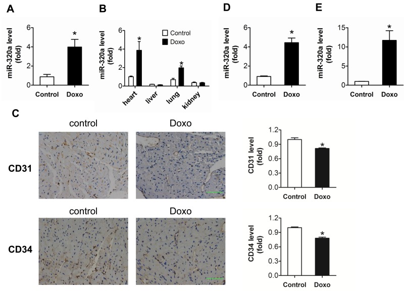
Background: Vascular homeostasis abnormalities may involve in doxorubicin induced cardiotoxicity.

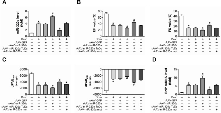
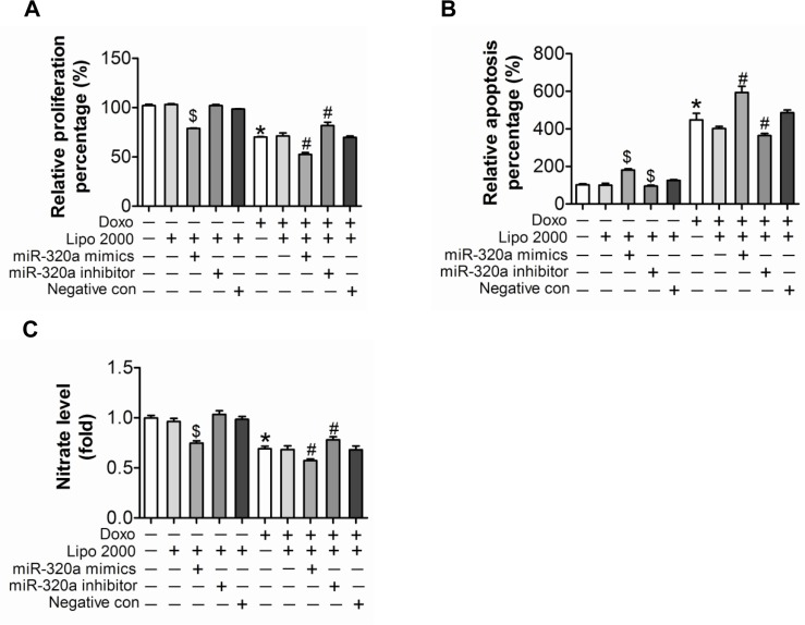
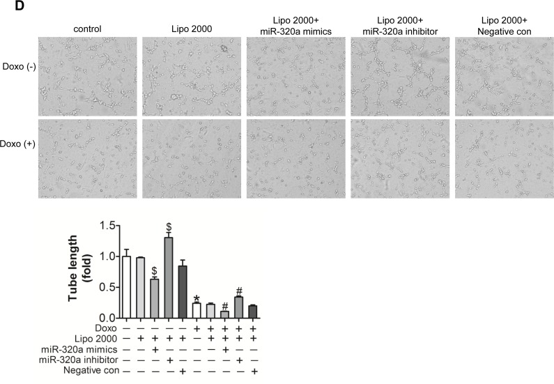
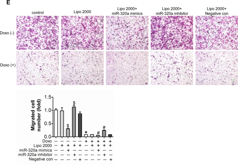
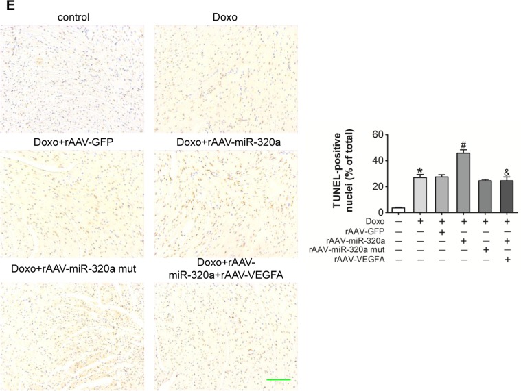
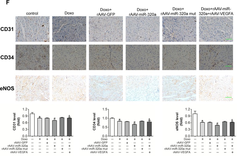
Methods: Enhanced cardiac miR-320a expression, reduced cardiac microvessel density and impaired cardiac function were observed in mice treated by anthracycline doxorubicin. To further explore the role of miR-320a in doxorubicin induced cardiotoxicity, microRNA mimics/inhibitor in vitro and rAAV administration in vivo were employed in mice.

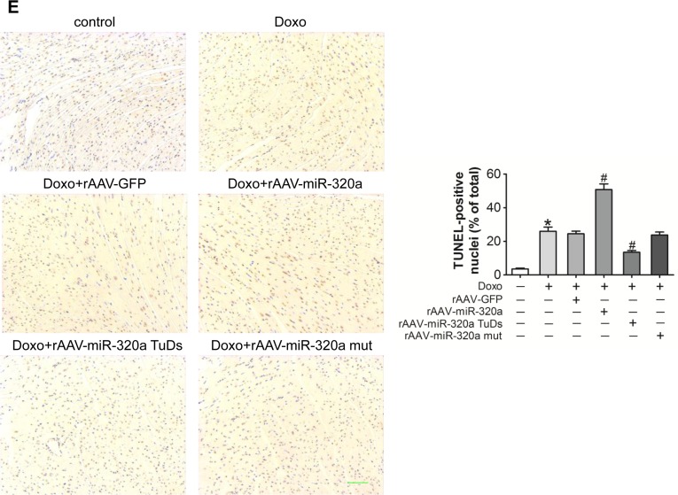
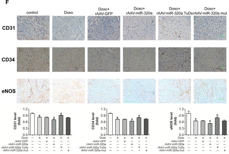
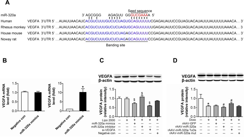
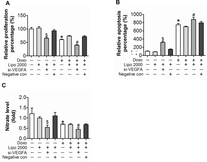
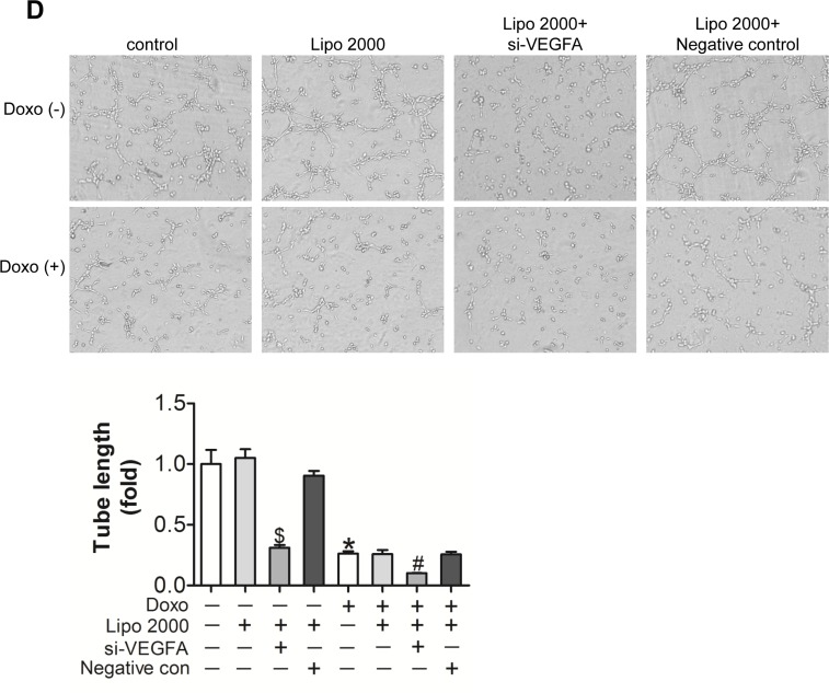
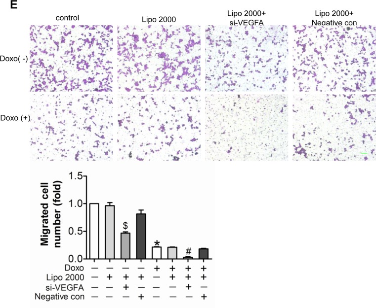
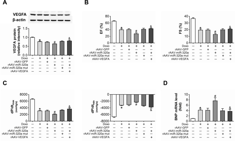
Results: Knockdown of miR-320a not only resulted in enhanced proliferation and inhibited apoptosis in cultured endothelial cells, but also attenuated cardiac abnormalities induced by doxorubicin. On the contrary, overexpression of miR-320a enhanced apoptosis in vitro, and aggravated vessel abnormalities in heart and subsequent cardiac dysfunction in mice. Furthermore, Western blot assays showed that VEGF-A was a potential target of miR-320a, which was verified by anti-Ago2 co-immunoprecipitation. Moreover, as same as miR-320a, siRNA against VEGF-A reinforced doxorubicin induced endothelial cells injury. Finally, the negative effects of miR-320a on vascular homeostasis and cardiac function were alleviated by VEGF-A re-expression in doxorubicin treated mice.

Conclusion: Our observations demonstrate that miR-320a play important roles in doxorubicin induced cardiotoxicity via vessel homeostasis in heart and thus, inhibition of miR-320a may be applied to the treatment of cardiac dysfunction induced by anthracycline.

Keywords: VEGF-A; cardiotoxicity; doxorubicin; miR-320a; vascular homeostasis.


Conflict of interest statement

Conflict of interest statement

The authors declare that they have no competing interests.

Figures

Figure 1
Figure 1. Increased miR-320a and decreased cardiac microvessel density were induced by doxorubicin in vivo and in vitro
(A) Relative cardiac miR-320a expression level measured by real-time PCR. (B) Relative expression of miR-320a among different organs measured by real-time PCR. (C) Expression level of CD31 and CD34 in heart detected by immunohistochemical staining. Scale bar, 200μm. (D) Relative miR-320a expression level in H9c2 cells measured by real-time PCR. (E) Relative miR-320a expression level in HUVEC cells measured by real-time PCR. Data are representative of three experiments, n≥4. Data are expressed as mean ± SEM, *P<0.05 versus control.
Figure 2
Figure 2. Inhibition of miR-320a improved cardiac dysfunction and cardiac microvessel injury induced by doxorubicin
(A) Cardiac expression of miR-320a detected by real-time PCR. (B) Echocardiographic detection of mice with different treatments. (C) Hemodynamic analysis measured by Millar cardiac catheter system of mice with different treatment. (D) Relative BNP expression level in heart from mice with different treatments measured by real-time PCR. (E) TUNEL staining of heart sections from mice with different treatments. (F) Expression level of CD31, CD34 and eNOS in heart detected by immunohistochemical staining. Scale bar, 200μm. Data are expressed as mean ± SEM, n≥4, *P<0.05 versus control, #P<0.05 versus Doxo.
Figure 3
Figure 3. Inhibition of miR-320a improved doxorubicin induced endothelial cells impairment in vitro
(A) Proliferation detected by BrdU incorporation assays. (B) Apoptosis measured by Annexin V/PI flow cytometric analysis. (C) NO release detected by Nitric oxide colorimetric assays. (D) Tube formation determined on Matrigel. (E) Migration evaluated by transwell experiment. Scale bar, 200μm. Data are representative of three experiments. Data are expressed as mean ± SEM, n≥3, *P<0.05 versus control, #P<0.05 versus Doxo + negative con, $P<0.05 versus negative control.
Figure 4
Figure 4. VEGF-A is a target of miR-320a
(A) Schematic representation of the predicted target sites of miR-320a in the 3′ UTR of VEGF-A. The 3′ end of predicted binding site in human VEGF-A is labeled with blue, the crucial seed regions in 5′ end of miR-320a is labeled with red. (B) Expression of VEGF-A in the whole RNA (left) or the RNA of the anti-Ago co-IP (right) from the cell lysates. (C) VEGF-A protein level in treated HUVEC. (D) VEGF-A protein level in treated mice. Data are representative of three experiments. Data are expressed as mean ± SEM, n≥3, *P<0.05 versus control, #P<0.05 versus Doxo + negative con or Doxo + rAAV-miR-320a mut, $P<0.05 versus negative con.
Figure 5
Figure 5. Down-regulated expression of VEGF-A aggravated doxorubicin-induced impairment in HUVEC
(A) Proliferation determined by BrdU incorporation assays. (B) Apoptosis measured by Annexin V/PI flow cytometric analysis. (C) NO release detected by Nitric oxide colorimetric assays. (D) Tube formation determined on Matrigel. (E) Migration evaluated by transwell experiment. Scale bar, 200μm. Data are representative of three experiments. Data are expressed as mean ± SEM, n≥3, *P<0.05 versus control, #P<0.05 versus Doxo + negative con, $P<0.05 versus negative con.
Figure 6
Figure 6. Restored VEGF-A eliminated the miR-320a induced cardiac dysfunction in doxorubicin treated mice
(A) Cardiac expression of VEGF-A detected by western blot analysis. (B) Echocardiographic detection of mice with different treatments. (C) Hemodynamic analysis measured by Millar cardiac catheter system of mice with different treatments. (D) Relative BNP expression level in heart measured by real-time PCR. (E) TUNEL staining in heart sections. (F) Expression level of CD31, CD34 and eNOS in heart detected by immunohistochemical staining. Scale bar, 200μm. Data are expressed as mean ± SEM, n≥4, *P<0.05 versus control, #P<0.05 versus Doxo + rAAV-miR-320a mut, $P<0.05 versus rAAV-miR-320a.

Similar articles

Cited by

References

    1. Carvalho FS, Burgeiro A, Garcia R, Moreno AJ, Carvalho RA, Oliveira PJ. Doxorubicin-induced cardiotoxicity: from bioenergetic failure and cell death to cardiomyopathy. Med Res Rev. 2014;34:106–135. - PubMed
    1. Yeh ET, Bickford CL. Cardiovascular complications of cancer therapy: incidence, pathogenesis, diagnosis, and management. Journal of the American College of Cardiology. 2009;53:2231–2247. - PubMed
    1. Cardinale D, Colombo A, Bacchiani G, Tedeschi I, Meroni CA, Veglia F, Civelli M, Lamantia G, Colombo N, Curigliano G, Fiorentini C, Cipolla CM. Early detection of anthracycline cardiotoxicity and improvement with heart failure therapy. Circulation. 2015;131:1981–1988. - PubMed
    1. Vejpongsa P, Yeh ET. Prevention of anthracycline-induced cardiotoxicity: challenges and opportunities. Journal of the American College of Cardiology. 2014;64:938–945. - PubMed
    1. Flood EC, Hajjar KA. The annexin A2 system and vascular homeostasis. Vascul Pharmacol. 2011;54:59–67. - PMC - PubMed

Publication types

MeSH terms

Substances