. 2016 Apr 7;44(6):2859-72.
doi: 10.1093/nar/gkw032. Epub 2016 Feb 2.

Recurrent chimeric fusion RNAs in non-cancer tissues and cells

Affiliations

Recurrent chimeric fusion RNAs in non-cancer tissues and cells

Mihaela Babiceanu et al. Nucleic Acids Res. .

Abstract

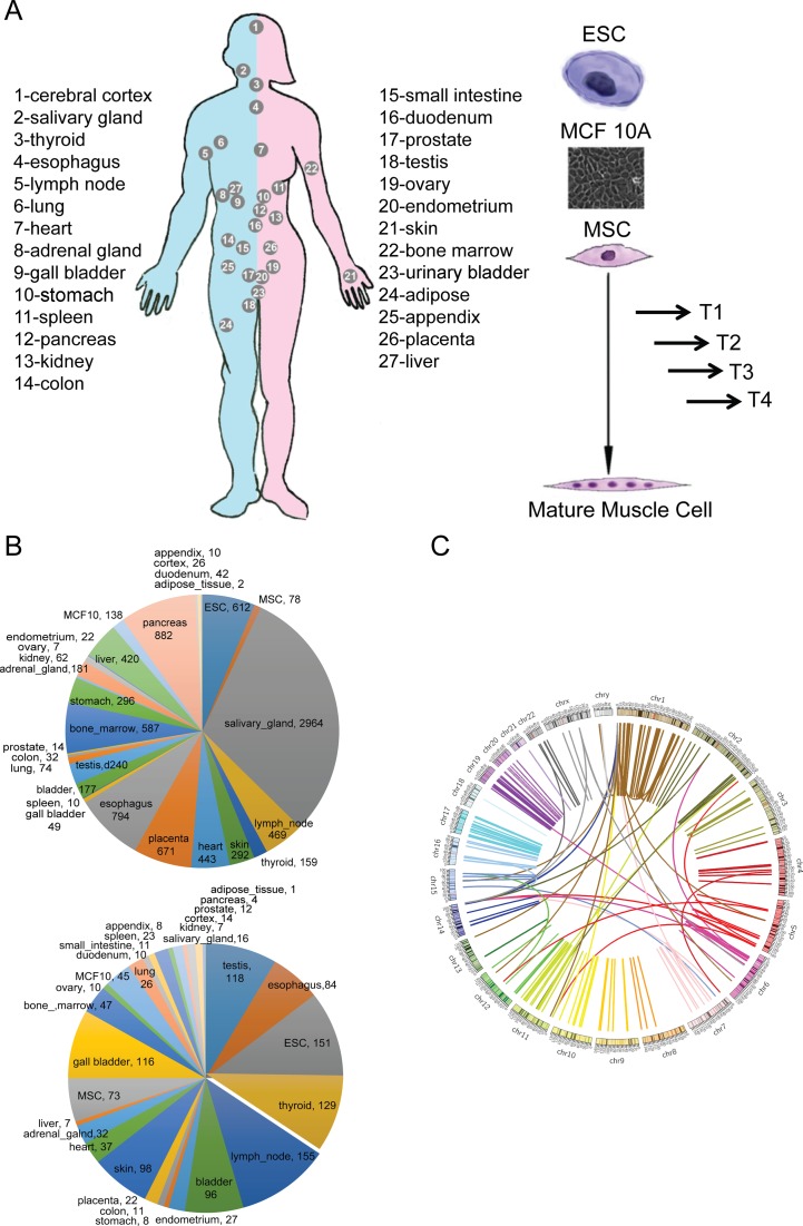
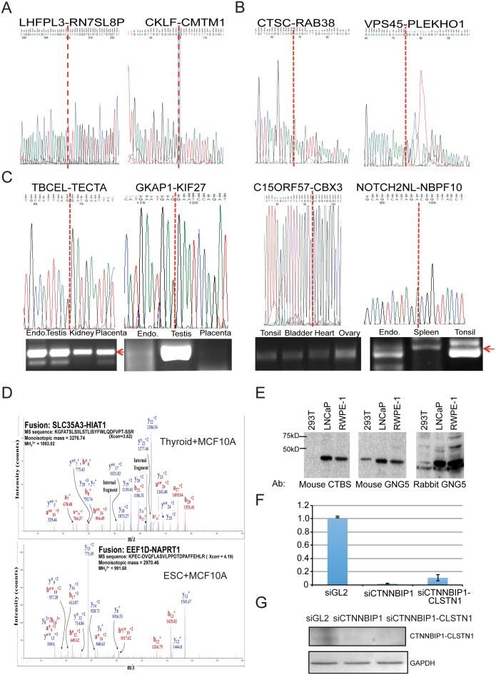
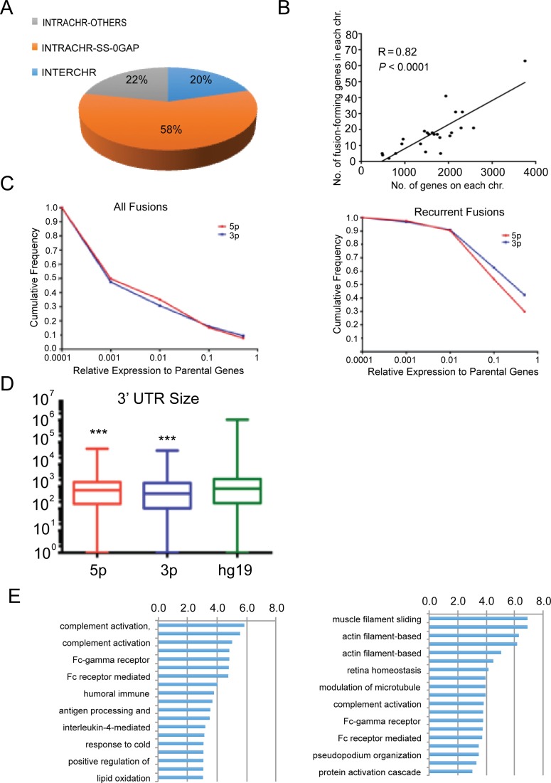
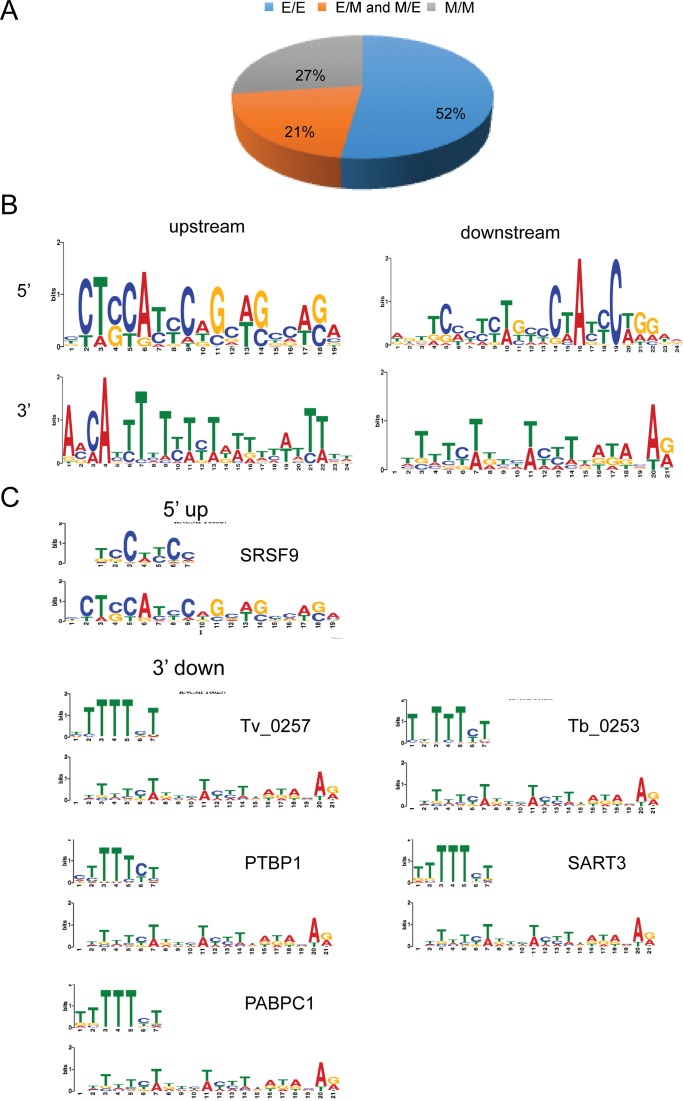
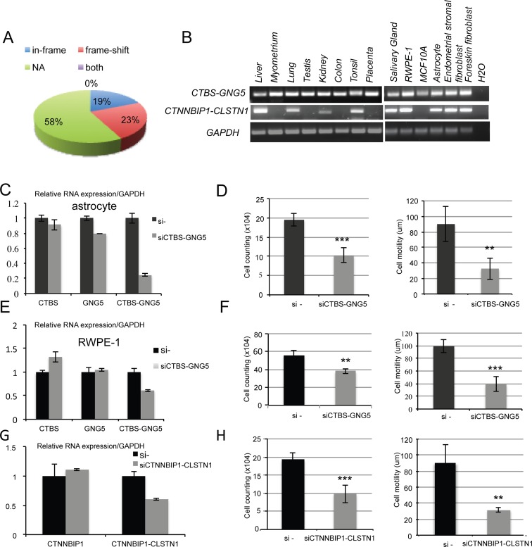
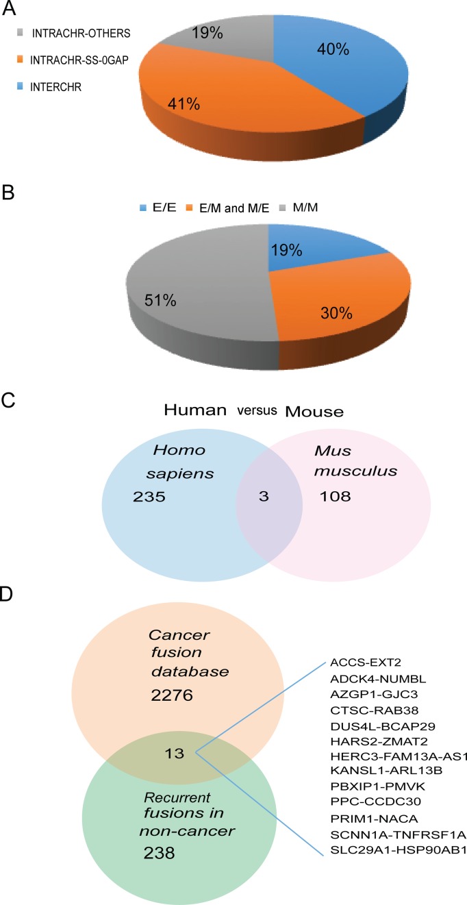
Gene fusions and their products (RNA and protein) were once thought to be unique features to cancer. However, chimeric RNAs can also be found in normal cells. Here, we performed, curated and analyzed nearly 300 RNA-Seq libraries covering 30 different non-neoplastic human tissues and cells as well as 15 mouse tissues. A large number of fusion transcripts were found. Most fusions were detected only once, while 291 were seen in more than one sample. We focused on the recurrent fusions and performed RNA and protein level validations on a subset. We characterized these fusions based on various features of the fusions, and their parental genes. They tend to be expressed at higher levels relative to their parental genes than the non-recurrent ones. Over half of the recurrent fusions involve neighboring genes transcribing in the same direction. A few sequence motifs were found enriched close to the fusion junction sites. We performed functional analyses on a few widely expressed fusions, and found that silencing them resulted in dramatic reduction in normal cell growth and/or motility. Most chimeras use canonical splicing sites, thus are likely products of 'intergenic splicing'. We also explored the implications of these non-pathological fusions in cancer and in evolution.


Figures

Figure 1.
Figure 1.
Identification of chimeric fusion RNAs in various tissues and cell types. (A) The sample sources include 27 different adult tissues on a human body map, ES cells, four time points collected along MSC muscle differentiation process, and MCF10A cells. (B) Distribution of fusions among different tissues/cells. Number of fusions in each sample type is also annotated. Upper: all the fusions; Lower: recurrent fusions. (C) Recurrent fusions were shown by a Circos plot. The fused genes are illustrated here as a line that connects two parental genes.
Figure 2.
Figure 2.
Validation of the fusions. (A) Sanger sequencing of RT-PCR products for two tissue-specific fusions. LHFPL3-RN7SL8P in lung (M/E) and CKLF-CMTM in testis (E/E). Red dotted lines indicate the fusion junction. (B) Sanger sequencing of RT-PCR product for two fusions identified during MSC muscle differentiation process. (C) RT-PCR and Sanger sequencing for four recurrent fusions. TBCEL-TECTA were detected in endometrium (endo.), testis, kidney and placenta (E/E, INTRACHR-SS-0GAP), GKAP1-KIF27 in endometrium, testis and placenta (E/E, INTRA-OTHERS), C15ORF57-CBX3 in tonsil, bladder, heart, ovary (E/M, INTERCHR) and NOTCH2NL-NBPF10 in endometrium, and tonsil (E/M, INTRACHR-SS-0GAP fusion). Red arrows point to the correct PCR product. (D) Two examples of chimeric peptides supported by LCMS in MCF10A cells. The superscripts ‘o’ and ‘*’ represent H2O and NH3 losses, respectively. (E) Western blot analyses using a CTBS antibody, and two GNG5 antibodies detecting CTBS-GNG5 protein. (F) qRT-PCR of CTNNBIP1-CLSTN1 normalized against GAPDH. Human foreskin fibroblast cells were transfected with si-negative control, siCTNNBIP1 and siCTNNBIP1-CLSTN1. (G) Western blot analyses of protein extracts from the same three samples as above. Upper: CLSTN1 antibody. Lower: GAPDH antibody.
Figure 3.
Figure 3.
Characteristics of the recurrent fusions. (A) Distribution of fusions according to the chromosomal location of the parental genes. INTERCHR: fusions involving parental genes located on different chromosomes; INTRACHR-SS-0GAP: fusions involving neighboring genes transcribing the same strand; and INTRACHR-OTHER: other fusions with parental genes on the same chromosome. (B) The density of genes participating in fusion formation correlates to the overall gene density on individual chromosomes. (C) Cumulative frequency of the relative expression of the fusion transcripts to their parental genes. Left: all the candidate fusions. Right: recurrent fusions. The parental gene expression was based on FPKM. The fusion RNA expression was converted into FPKM from the sum of junction and split reads number. (D) Box plot depicting the comparison between the 3′ UTR size for the parental 5′ and 3′ genes involved in fusions, and all genes known to date in hg19. (E) Gene ontology terms enriched in 5′ parental genes and 3′ parental genes involved in recurrent fusions. Plotted are statistical significance (-Log10(P value)) of each term.
Figure 4.
Figure 4.
Further characterization of the recurrent fusions. (A) Distribution of fusions according to the junction position relative to the parental exons. E/E: both 5′ and 3′ using known exon boundaries; E/M or M/E: one side using known exon boundary, the other not; M/M: both sides fall into the middle of known exons. (B) Motif scanning using sequences 200 bp immediately upstream or downstream to the fusion junction site of both 5′ genes and 3′ genes. Shown are the motifs found having the highest GLAM2 scores. (C) Known RNA-binding motifs matching the motifs found through MEME. Using P = 0.001 as cutoff, one known RNA-binding motif was found similar to the 5′ gene downstream motif. Five RNA-binding motifs were found similar to the 3′ gene downstream motif.
Figure 5.
Figure 5.
Functional relevance of the fusions. (A) Distribution of the fusions according to their protein-coding potential: the known protein coding sequence of the 3′ gene uses a different reading frame than the 5′ gene (frame-shift); the known reading frame of the 3′ gene is the same as the 5′ gene (in-frame); no effect on the reading frame of the parental genes (NA). (B) CTBS-GNG5 and CTNNBIP1-CLSTN1 are widely expressed among tissues and cell lines. (C) An siRNA targeting CTBS-GNG5 resulted in significant reduction of the fusion transcript in immortalized astrocytes. (D) Knocking down CTBS-GNG5 using the fusion-targeting siRNA resulted in significance growth suppression and reduction in cell motility. (E) The siRNA also specifically silenced CTBS-GNG5 fusion transcript in RWPE-1 cells. (F) Knocking down CTBS-GNG5 in RWPE cells also resulted in reduced cell growth and motility. (G) An siRNA targeting CTNNBIP1-CLSTN1 resulted in significant reduction of the fusion transcript, but not wild-type CTNNBIP1 in immortalized astrocytes. Wild-type CLSTN1 was undetectable in these cells. (H) Knocking down CTNNBIP1-CLSTN1 in these astrocyte cells resulted in reduced cell growth and motility. **P < 0.005. ***P < 0.001.
Figure 6.
Figure 6.
Conservation and cancer implications. (A) Fusion RNAs of 81 samples from 15 mouse tissues. Similar to that in human, INTRACHR-SS-0GAP also constituted the biggest portion. (B) Distribution of fusions according to the junction position relative to the parental exon. (C) Venn diagram showing the similarities/differences of fusions in Homo sapiens versus Mus musculus. (D) Venn diagram showing the common fusions in the normal tissues/cells versus the Mitelman Database of Chromosome Aberrations and Gene Fusions in Cancer. Thirteen common fusions are listed.

References

    1. Rowley J.D. The role of chromosome translocations in leukemogenesis. Semin. Hematol. 1999;36:59–72. - PubMed
    1. Heim S., Mitelman F. Molecular screening for new fusion genes in cancer. Nat. Genet. 2008;40:685–686. - PubMed
    1. Chase A., Ernst T., Fiebig A., Collins A., Grand F., Erben P., Reiter A., Schreiber S., Cross N.C. TFG, a target of chromosome translocations in lymphoma and soft tissue tumors, fuses to GPR128 in healthy individuals. Haematologica. 2010;95:20–26. - PMC - PubMed
    1. Ren G., Zhang Y., Mao X., Liu X., Mercer E., Marzec J., Ding D., Jiao Y., Qiu Q., Sun Y., et al. Transcription-mediated chimeric RNAs in prostate cancer: time to revisit old hypothesis. OMICS. 2014;18:615–624. - PMC - PubMed
    1. Yoshihara K., Wang Q., Torres-Garcia W., Zheng S., Vegesna R., Kim H., Verhaak R.G. The landscape and therapeutic relevance of cancer-associated transcript fusions. Oncogene. 2015;34:4845–4854. - PMC - PubMed

Publication types

MeSH terms