

Organization of descending neurons in Drosophila melanogaster
- PMID: 26837716
- PMCID: PMC4738306
- DOI: 10.1038/srep20259
Organization of descending neurons in Drosophila melanogaster
Abstract
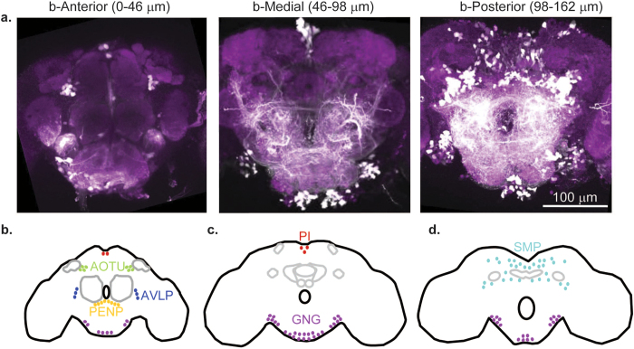
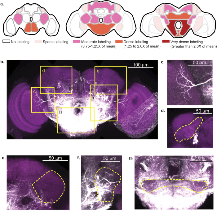
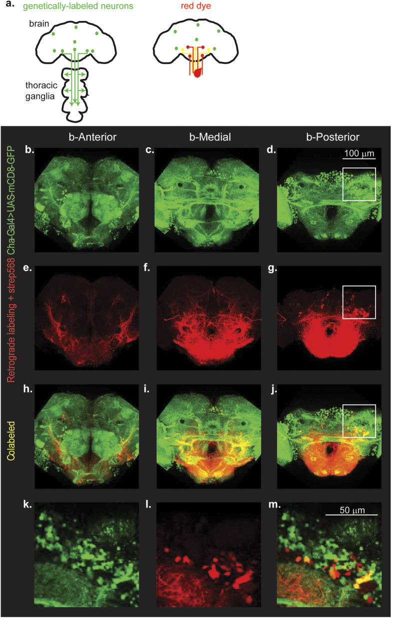
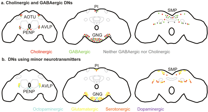
Neural processing in the brain controls behavior through descending neurons (DNs) - neurons which carry signals from the brain to the spinal cord (or thoracic ganglia in insects). Because DNs arise from multiple circuits in the brain, the numerical simplicity and availability of genetic tools make Drosophila a tractable model for understanding descending motor control. As a first step towards a comprehensive study of descending motor control, here we estimate the number and distribution of DNs in the Drosophila brain. We labeled DNs by backfilling them with dextran dye applied to the neck connective and estimated that there are ~1100 DNs distributed in 6 clusters in Drosophila. To assess the distribution of DNs by neurotransmitters, we labeled DNs in flies in which neurons expressing the major neurotransmitters were also labeled. We found DNs belonging to every neurotransmitter class we tested: acetylcholine, GABA, glutamate, serotonin, dopamine and octopamine. Both the major excitatory neurotransmitter (acetylcholine) and the major inhibitory neurotransmitter (GABA) are employed equally; this stands in contrast to vertebrate DNs which are predominantly excitatory. By comparing the distribution of DNs in Drosophila to those reported previously in other insects, we conclude that the organization of DNs in insects is highly conserved.
Conflict of interest statement
The authors declare no competing financial interests.
Figures
Similar articles
-
Vesicular neurotransmitter transporters in Drosophila melanogaster.Biochim Biophys Acta Biomembr. 2020 Dec 1;1862(12):183308. doi: 10.1016/j.bbamem.2020.183308. Epub 2020 Apr 17. Biochim Biophys Acta Biomembr. 2020. PMID: 32305263 Free PMC article. Review.
-
Chemical neuroanatomy of the Drosophila central complex: distribution of multiple neuropeptides in relation to neurotransmitters.J Comp Neurol. 2011 Feb 1;519(2):290-315. doi: 10.1002/cne.22520. J Comp Neurol. 2011. PMID: 21165976
-
The functional organization of descending sensory-motor pathways in Drosophila.Elife. 2018 Jun 26;7:e34272. doi: 10.7554/eLife.34272. Elife. 2018. PMID: 29943730 Free PMC article.
-
Neurotransmitter-induced changes in the intracellular calcium concentration suggest a differential central modulation of CCAP neuron subsets in Drosophila.Dev Neurobiol. 2007 May;67(6):792-808. doi: 10.1002/dneu.20392. Dev Neurobiol. 2007. PMID: 17443825
-
Lessons from sleeping flies: insights from Drosophila melanogaster on the neuronal circuitry and importance of sleep.J Neurogenet. 2013 Jun;27(1-2):23-42. doi: 10.3109/01677063.2013.791692. Epub 2013 May 23. J Neurogenet. 2013. PMID: 23701413 Review.
Cited by
-
IRF2BPL Is Associated with Neurological Phenotypes.Am J Hum Genet. 2018 Aug 2;103(2):245-260. doi: 10.1016/j.ajhg.2018.07.006. Epub 2018 Jul 26. Am J Hum Genet. 2018. PMID: 30057031 Free PMC article.
-
On the Role of the Head Ganglia in Posture and Walking in Insects.Front Physiol. 2020 Feb 21;11:135. doi: 10.3389/fphys.2020.00135. eCollection 2020. Front Physiol. 2020. PMID: 32153430 Free PMC article. Review.
-
Transgenic line for characterizing GABA-receptor expression to study the neural basis of olfaction in the yellow-fever mosquito.Front Physiol. 2024 Mar 28;15:1381164. doi: 10.3389/fphys.2024.1381164. eCollection 2024. Front Physiol. 2024. PMID: 38606012 Free PMC article.
-
Descending control of motor sequences in Drosophila.Curr Opin Neurobiol. 2024 Feb;84:102822. doi: 10.1016/j.conb.2023.102822. Epub 2023 Dec 13. Curr Opin Neurobiol. 2024. PMID: 38096757 Free PMC article. Review.
-
The role of learning-walk related multisensory experience in rewiring visual circuits in the desert ant brain.J Comp Physiol A Neuroethol Sens Neural Behav Physiol. 2023 Jul;209(4):605-623. doi: 10.1007/s00359-022-01600-y. Epub 2022 Dec 9. J Comp Physiol A Neuroethol Sens Neural Behav Physiol. 2023. PMID: 36494572 Free PMC article. Review.
References
-
- Staudacher Y. E. Gating of sensory responses of descending brain neurones during walking in crickets. The Journal of experimental biology 201 (Pt 4), 559–572 (1998). - PubMed
Publication types
MeSH terms
Substances
LinkOut - more resources
Full Text Sources
Other Literature Sources
Molecular Biology Databases
Research Materials
