Review
. 2016 Apr;73(8):1623-40.
doi: 10.1007/s00018-016-2142-0. Epub 2016 Feb 2.

Aquaporin-5: from structure to function and dysfunction in cancer

Affiliations
Review

Aquaporin-5: from structure to function and dysfunction in cancer

Inês Direito et al. Cell Mol Life Sci. 2016 Apr.

Abstract

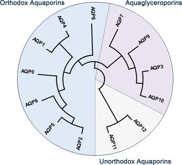
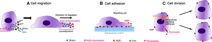
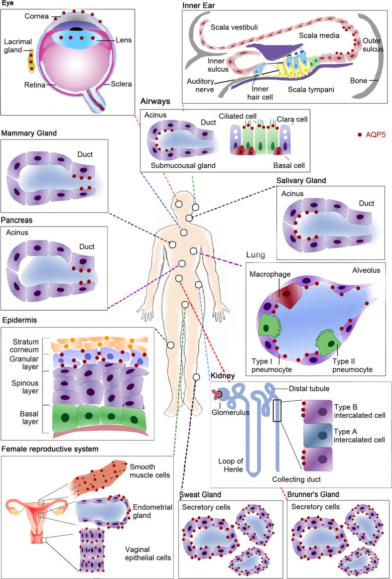
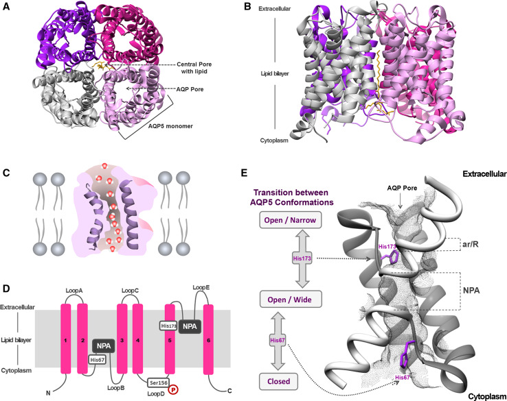
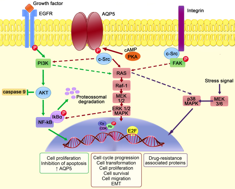
Aquaporins, a highly conserved group of membrane proteins, are involved in the bidirectional transfer of water and small solutes across cell membranes taking part in many biological functions all over the human body. In view of the wide range of cancer malignancies in which aquaporin-5 (AQP5) has been detected, an increasing interest in its implication in carcinogenesis has emerged. Recent publications suggest that this isoform may enhance cancer cell proliferation, migration and survival in a variety of malignancies, with strong evidences pointing to AQP5 as a promising drug target and as a novel biomarker for cancer aggressiveness with high translational potential for therapeutics and diagnostics. This review addresses the structural and functional features of AQP5, detailing its tissue distribution and functions in human body, its expression pattern in a variety of tumors, and highlighting the underlying mechanisms involved in carcinogenesis. Finally, the actual progress of AQP5 research, implications in cancer biology and potential for cancer detection and prognosis are discussed.

Keywords: Aquaporin; Biomarker; Permeability; Signaling pathways; Tumor; Water channel.


Figures

Fig. 1
Fig. 1
Dendrogram depicting the phylogenetic relationship of mammalian aquaporins (AQP), showing the three main subfamilies. Dendogram was generated by neighbor-joining method (applied to 1000 bootstrap data sets) using the MEGA6 software [145]. The accession numbers are as follows: MIP (XP_011536656), AQP1 (NP_932766), AQP2 (NP_000477), AQP3 (NP_004916), AQP4 (NP_001641), AQP5 (NP_001642), AQP6 (NP_001643), AQP7 (NP_001161), AQP8 (NP_001160), AQP9 (NP_066190), AQP10 (NP_536354), AQP11 (NP_766627), AQP12 (NP_945349)
Fig. 2
Fig. 2
Aquaporins (AQPs) roles in cancer. a At the leading edge of the migrating cell, AQPs facilitate water fluxes driven by an increase in local osmolarity due to transmembrane ion fluxes, promoting lamellipodium formation and stabilization by actin polymerization. b AQPs may increase cell–matrix adhesion, important for tumor spread. c AQPs facilitate permeants (glycerol, hydrogen peroxide) uptake or may interact with oncoproteins, which activate intracellular signaling cascades promoting the transcription of genes involved in tumor cell proliferation
Fig. 3
Fig. 3
Tissue distribution of human aquaporin-5 (AQP5). In the eye AQP5 is expressed in the cytoplasm and/or plasma membrane of lens fibber cells depending on its differentiation, in the cytoplasm and plasma membrane of corneal epithelium and in the apical membrane of acinar cells in lacrimal glands. In the inner ear this isoform is expressed in the cochlea, in the apical membrane of outer sulcus cells of the apical turn. In the respiratory system AQP5 is expressed in airways at the apical membrane of columnar epithelial cells and at the apical membrane of serous acinar cells of sub-mucosal glands. In lungs, AQP5 is expressed at the apical membrane of type I pneumocytes. It is also expressed in the apical membrane of ductal cells in mammary glands. In the digestive system this isoform is expressed in the pancreas, at the apical membrane of intercalated and intralobular ductal cells and at the apical membrane of acinar cells in salivary glands, as well as at the apical and basolateral membranes of secretory cells of Brunner’s glands in the duodenum. In integumentary system AQP5 is expressed in the plasma membrane of keratinocytes in the skin granular layer and in apical and basolateral membranes of secretory cells in sweat glands. AQP5 is also expressed in renal cortex, in the apical membrane of type B intercalated cells in the collecting duct. In the female reproductive system AQP5 is expressed in the cytoplasm of vaginal epithelial cells, in the basolateral membrane of endometrial glandular epithelial cells and in the plasma membrane of uterus smooth muscle cells
Fig. 4
Fig. 4
Structure of AQP5. a Extracellular and b side views of AQP5 homotetramer showing the central pore with lipid (phosphatidylserine). c Diagram illustrating how water molecules permeate through AQP pore. d Topology map of the basic monomeric AQP5 fold, showing the six transmembrane alpha-helices (16) connected by loops (AE), the conserved asparagine–proline–alanine (NPA) motifs embed in the membrane, the histidine residues involved in gating (His67 and His173) and the serine residue involved in intracellular signaling (Ser156). e Detailed view of AQP5 pore and schematic representation of the proposed AQP5 gating mechanism. The two half-helices are depicted in white and the positioning of ar/R and NPA selectivity filters is indicated. The grey mesh represents the residues lining AQP5 pore. Key histidine residues involved in AQP5 gating are highlighted: His67, which controls the transition between closed and open conformations, and His173, controlling the transition between wide and narrow states. Structures were generated with Chimera (http://www.cgl.ucsf.edu/chimera) and are based on AQP5 X-ray structure (protein data bank code: 3D9S)
Fig. 5
Fig. 5
AQP5 intracellular signaling pathways involved in cancer. Phosphorylation of AQP5, mediated by cAMP-dependent protein kinase A (PKA), promotes the binding of adaptor molecules with SH3 domain, such as c-Src, triggering intracellular signaling cascades. Downstream, there is signal transduction through RAS/Raf-1/mitogen activated protein kinase 1 and 2 (MEK1/2)/extracellular signal-regulated kinases 1 and 2 (ERK1/2) pathway to the nucleus, where cyclin (Cy)/cyclin-dependent kinases (CDK) complexes phosphorylate retinoblastoma protein (Rb) that releases the transcription factor E2F, leading to expression of genes that are involved in cell transformation, proliferation, cycle progression and survival. Cell–matrix adhesion mediated by integrins is required for the continuous activation of focal adhesion kinase–mitogen-activated protein kinases (FAK–MAPK) pathway that is essential for cytoskeleton integrity. This pathway also promotes epithelial mesenchymal transition (EMT) by increasing the expression of mesenchymal markers and decreasing the expression of epithelial markers, essential for cancer cell migration and spread. Epidermal growth factor receptor (EGFR) activation by growth factors can activate RAS/MAPK signal transduction pathway and phosphoinositide 3-kinase (PI3K), which in turn activate protein kinase B (AKT) that is able to block caspase-9 activation. In the cytoplasm, nuclear factor-κB (NF-κB) binds to its inhibitor IκBα and stays in an inactive state. After receptor activation, IκBα is phosphorylated and undergoes proteossomal degradation. NF-κB is released and translocates into the nucleus where it activates the transcription of target genes, namely anti-apoptotic genes, resulting in increased AQP5 expression, cell proliferation and survival. AQP5 expression was also recently implicated in colon cancer multidrug resistance mechanisms due to activation of p38 MAPK pathway in response to stress signals

References

    1. Paganelli CV, Solomon AK. The rate of exchange of tritiated water across the human red cell membrane. J Gen Physiol. 1957;41(2):259–277. doi: 10.1085/jgp.41.2.259. - DOI - PMC - PubMed
    1. Whittembury G. Ion and water transport in the proximal tubules of the kidney of Necturus maculosus. J Gen Physiol. 1960;43:43–56. doi: 10.1085/jgp.43.5.43. - DOI - PMC - PubMed
    1. Smith BL, Agre P. Erythrocyte Mr 28,000 transmembrane protein exists as a multisubunit oligomer similar to channel proteins. J Biol Chem. 1991;266(10):6407–6415. - PubMed
    1. Agre P, Saboori AM, Asimos A, Smith BL. Purification and partial characterization of the Mr 30,000 integral membrane protein associated with the erythrocyte Rh(D) antigen. J Biol Chem. 1987;262(36):17497–17503. - PubMed
    1. Preston GM, Agre P. Isolation of the cDNA for erythrocyte integral membrane protein of 28 kilodaltons: member of an ancient channel family. Proc Natl Acad Sci USA. 1991;88(24):11110–11114. doi: 10.1073/pnas.88.24.11110. - DOI - PMC - PubMed

Publication types

LinkOut - more resources