. 2016 Feb 2:14:36.
doi: 10.1186/s12967-016-0789-9.

Colorectal cancer-derived microvesicles modulate differentiation of human monocytes to macrophages

Affiliations

Colorectal cancer-derived microvesicles modulate differentiation of human monocytes to macrophages

Monika Baj-Krzyworzeka et al. J Transl Med. .

Abstract

Background: Tumour-derived microvesicles (TMVs) are important players in tumour progression, modulating biological activity of immune cells e.g. lymphocytes, monocytes and macrophages. This phenomenon is particularly interesting in the progression of colon cancer, as macrophages in this type of tumour are relevant for the recovery processes. In the present study, the role of colon cancer cell-derived microvesicles in monocyte differentiation and activity profile (polarization) was investigated.

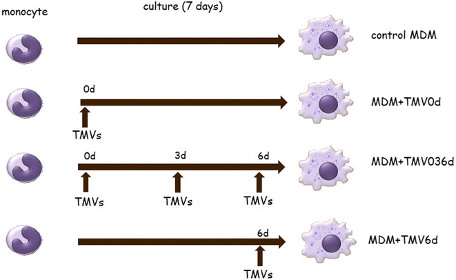
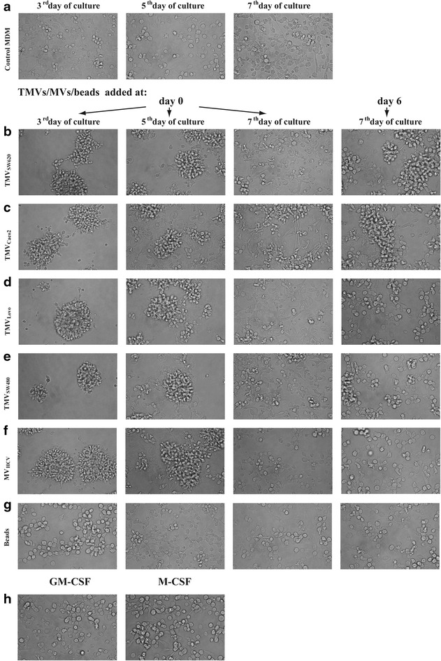
Methods: Monocyte-derived macrophages (MDM) were differentiated in vitro in the presence of TMVs obtained from colon cancer: Caco-2, SW620, LoVo or SW480 cell lines and analysed according to their morphology and biological functions, as defined by cytokine secretion, reactive oxygen intermediate (ROI) production and cytotoxic activity against respective colon cancer cells.

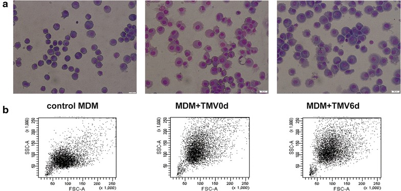
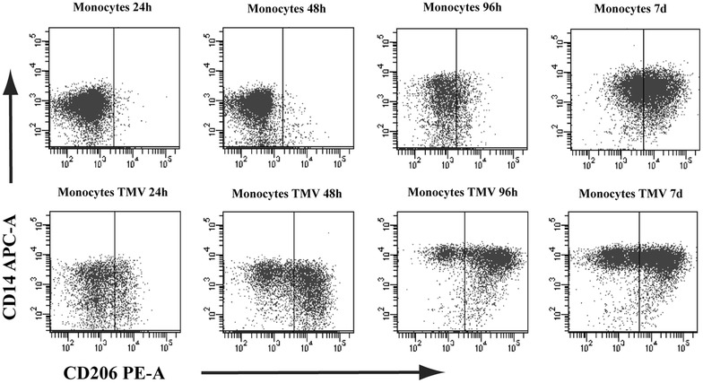
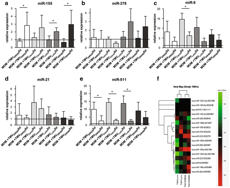
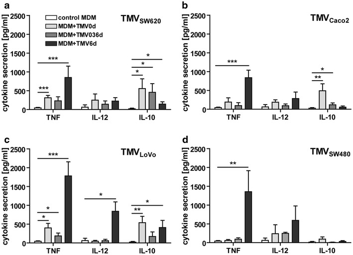
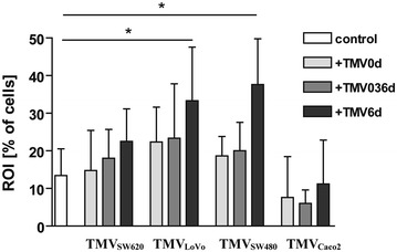
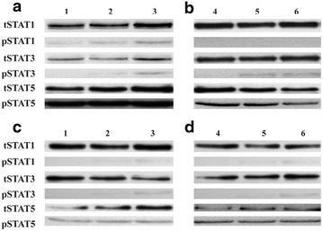
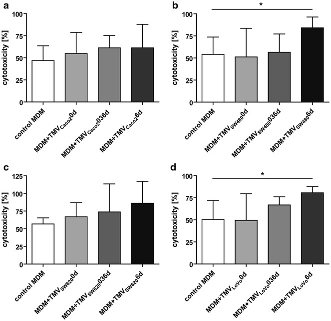
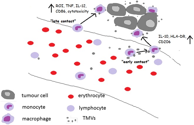
Results: Monocytes differentiated with TMVs exhibited morphological and phenotypical characteristics of macrophages. An early contact (beginning with the first day of the in vitro culture) of monocytes with TMVs resulted in increased IL-10 secretion and only slightly elevated TNF release. Early, or prolonged contact resulted in low ROI production and low cytotoxicity against tumour cells. On the other hand, late contact of MDM with TMVs, stimulated MDM to significant TNF and IL-12 secretion, ROI production and enhanced cytotoxicity against tumour cells in vitro. In addition, differences in MDM response to TMVs from different cell lines were observed (according to cytokine secretion, ROI production and cytotoxicity against tumour cells in vitro). Biological activity, STATs phosphorylation and microRNA profiling of MDMs indicated differences in their polarization/activation status which may suggest mixed polarization type M1/M2 with the predominance of proinflammatory cells after late contact with TMVs.

Conclusions: Macrophage activity (polarization status) may be regulated by contact with not only tumour cells but also with TMVs. Their final polarization status depends on the contact time, and probably on the vesicle "cargo", as signified by the distinct impact of TMVs which enabled the switching of MDM maturation to regulatory macrophages.


Figures

Fig. 1
Fig. 1
Scheme of experiments. Control MDM were obtained from human peripheral blood monocytes cultured for 7 days without growth factors. MDM + TMV0d were obtained from human peripheral blood monocytes supplemented at day 0 with appropriate TMVs. Similarly, MDM + TMV036d were exposed to TMVs at day 0, 3 and 6 and MDM + TMV6d only at day 6. All other culture conditions were the same
Fig. 2
Fig. 2
Morphology of MDM (at day 3, 5 and 7) differentiated in the presence of medium alone (a) or TMVs: TMVSW620 (b), TMVCaco2 (c), TMVLoVo (d), TMVSW480 (e), MVHCV (f), beads (g) and growth factors (h). One representative experiment out of ten is presented
Fig. 3
Fig. 3
MDM morphology analysed by cytospin slides after the Wright’s staining (a) and flow cytometry FSC/SSC dot plot (b) of MDM differentiated in the presence of TMVs. Left panel-control, middle panel-MDM + TMV0d, right panel-MDM + TMV6d. (bar—50 µm). One representative example out of ten is presented
Fig. 4
Fig. 4
Expression of CD206 on MDM cultured alone (upper panel) or with TMVLoVo0d (lower panel) after 24, 48, 96 h and 7 days. One representative experiment out of 6 performed is presented. Similar kinetics of CD206 expression was observed for other TMVs
Fig. 5
Fig. 5
Expression of selected (involved in the MDM differentiation process) microRNAs in MDM + TMV0d and MDM + TMV6d vs control MDM (black line at level 1) presented as relative expression normalized to U6 (2−ΔΔCT): miR-155 (a), miR-378 (b), miR-9 (c), miR-21 (d), miR-511 (e). Heat map of microRNA involved in MDM differentiation process detected in TMVs alone (f). Data from 3 independent experiments, performed in triplicates (mean ± SD) are shown. *p < 0.05
Fig. 6
Fig. 6
Secretion of cytokines (TNF, IL-12, IL-10) by MDM differentiated with TMVs. The supernatants were collected at day 7 and cytokines level was determined by ELISA method. Cytokine secretion by MDM culture with TMVSW620 (a), TMVCaco2 (b), TMVLoVo (c), TMVSW480 (d) is presented. Data from 6 independent experiments (mean ± SD) are shown. *p < 0.05, **p < 0.001, ***p < 0.0001
Fig. 7
Fig. 7
The intracellular production of ROI by MDM differentiated in the presence of TMVs. The level of ROI (mainly O2 ) production was determined by flow cytometry. MDM were stimulated with PMA in the presence of HE for 30 min. Percentage of positive cells was presented (mean of six performed experiments). Data from 6 independent experiments (mean ± SD) are shown. *p < 0.05
Fig. 8
Fig. 8
Western-blot analysis of STAT1, 3 and 5 phosphorylation. Monocytes isolated from blood of 2 donors were used: donor 1 (a and c), donor 2 (b and d). a and b represents MDM0d, c and d represents MDM6d. Control MDM (1 and 4), MDM + TMVLoVo (2), MDM + TMVSW480 (3), MDM + TMVSW620 (5) MDM + TMVCaco2 (6). One representative experiment out of two performed is presented
Fig. 9
Fig. 9
Cytotoxic/cytostatic activity of MDM against tumour cells. Monocytes were differentiated in the presence of TMVs and then cocultured with appropriate tumour cells for 48 h (a Caco-2, b SW480, c SW620, d LoVo). Proliferation of cells was determined by MTT reduction assay. The results of five independent experiments are shown (mean ± SD). *p < 0.05
Fig. 10
Fig. 10
Presentation of the hypothetical interactions of monocytes/macrophages with TMVs in the blood and in the tumour bed. Early contact of monocytes with TMVs resulting in M1/M2 mix polarization is schematically presented on the right. Late contact of macrophages with TMVs resulting in the induction of proinflammatory cells—left side. TMVs interactions with other immune cells as well as with tumour cells were omitted to simplify the scheme

References

    1. Weitz J, Koch M, Debus J, Hohler T, Galle PR, Buchler MW. Colorectal cancer. Lancet. 2005;14:153–165. doi: 10.1016/S0140-6736(05)17706-X. - DOI - PubMed
    1. Swiderska M, Choromańska B, Dąbrowska E, Konarzewska-Duchnowska E, Choromańska K, Szczurko G, Myśliwiec P, Dadan J, Ladny JR, Zwierz K. The diagnostics of colorectal cancer. Contemp Oncol (Pozn) 2014;18:1–6. - PMC - PubMed
    1. Brenner H, Kloor M, Pox CP. Colorectal cancer. Lancet. 2014;383:1490–1502. doi: 10.1016/S0140-6736(13)61649-9. - DOI - PubMed
    1. Mantovani A, Germano G, Marchesi F, Locatelli M, Biswas SK. Cancer-promoting tumour-associated macrophages: new vistas and open questions. Eur J Immunol. 2011;41:2522–2525. doi: 10.1002/eji.201141894. - DOI - PubMed
    1. Mantovani A, Sozzani S, Locati M, Allavena P, Sica A. Macrophage polarization: tumour-associated macrophages as a paradigm for polarized M2 mononuclear phagocytes. Trends Immunol. 2002;23:549–555. doi: 10.1016/S1471-4906(02)02302-5. - DOI - PubMed

Publication types

MeSH terms