

Hyperintense perilesional edema in the brain on T1-weighted images: Cavernous malformation or metastatic melanoma? Three case reports and literature review
- PMID: 26838172
- PMCID: PMC4978347
- DOI: 10.1177/1971400915626430
Hyperintense perilesional edema in the brain on T1-weighted images: Cavernous malformation or metastatic melanoma? Three case reports and literature review
Abstract
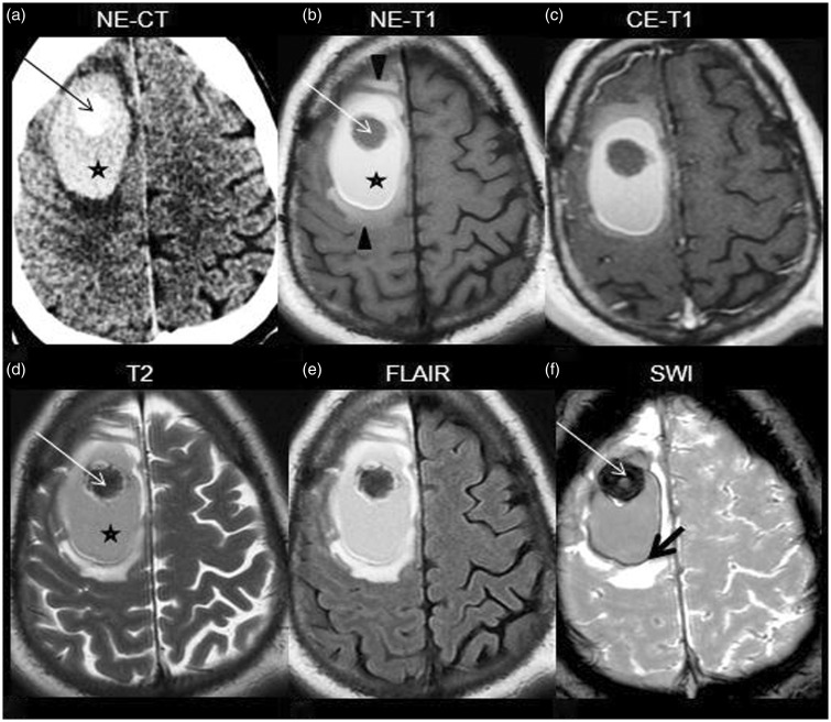
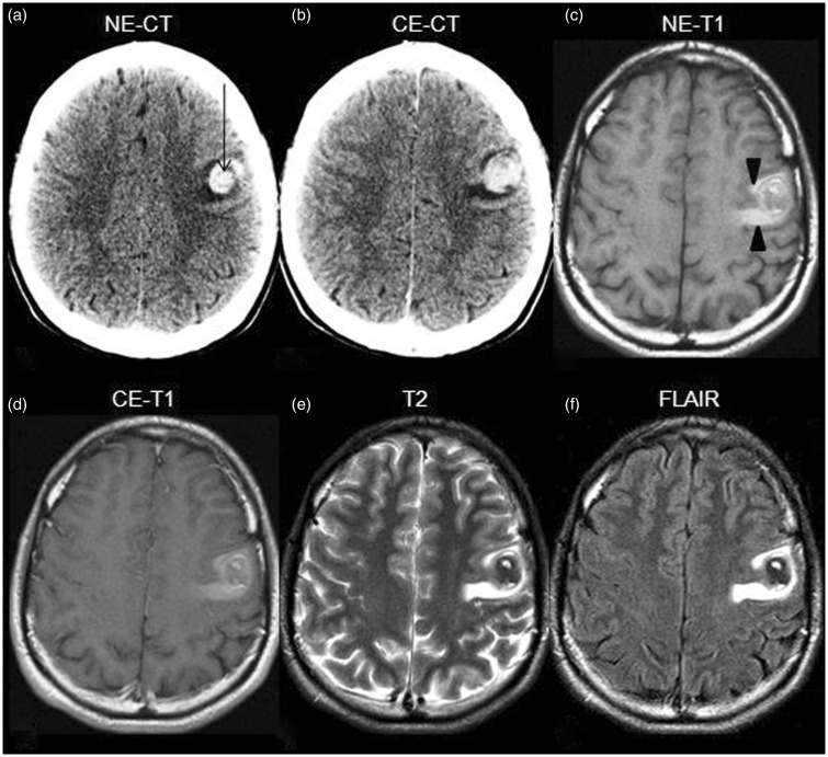
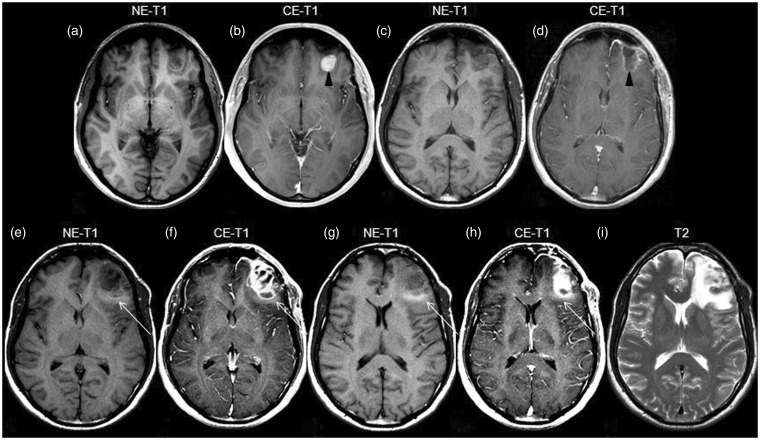
Hyperintense perilesional edema in brain masses on T1-weighted images (T1WI) is an unusual radiological finding. We report three cases showing this particular type of edema, one representing cerebral hemorrhagic cavernous malformation (CCM, cavernoma) and the other two, metastases of melanoma. The association between this sign and cavernoma was recently recognized. On the other hand, in melanotic lesions, the relationship with T1WI-hyperintense perilesional edema has not yet been described. Despite being an infrequent sign, it can considerably narrow the differential diagnosis, which gives it a high value for clinical practice. Moreover, given the high prevalence of the entities that manifest this imaging feature, it can be occasionally noticed.
Keywords: Cerebral cavernous malformation; cavernoma; hyperintense edema on T1; melanoma.
© The Author(s) 2016.
Figures
Similar articles
-
Hyperintense Perilesional Edema on T1-Weighted Imaging in Cavernoma: A Case Report.Cureus. 2024 Jan 1;16(1):e51454. doi: 10.7759/cureus.51454. eCollection 2024 Jan. Cureus. 2024. PMID: 38169697 Free PMC article.
-
Perilesional Hyperintensity on T1-Weighted Images in Intra-Axial Brain Masses other than Cavernous Malformations.J Neuroimaging. 2017 Sep;27(5):531-538. doi: 10.1111/jon.12424. Epub 2017 Jan 18. J Neuroimaging. 2017. PMID: 28098958
-
A T1 hyperintense perilesional signal aids in the differentiation of a cavernous angioma from other hemorrhagic masses.AJNR Am J Neuroradiol. 2008 Mar;29(3):494-500. doi: 10.3174/ajnr.A0847. Epub 2007 Nov 26. AJNR Am J Neuroradiol. 2008. PMID: 18039756 Free PMC article.
-
A peri-trigonal giant tumefactive cavernous malformation: case report and review of literature.Childs Nerv Syst. 2010 Dec;26(12):1819-23. doi: 10.1007/s00381-010-1237-4. Epub 2010 Jul 28. Childs Nerv Syst. 2010. PMID: 20665038 Review.
-
Giant cerebral cavernoma. Case report with literature review.Neurosciences (Riyadh). 2012 Jan;17(1):69-73. Neurosciences (Riyadh). 2012. PMID: 22246016 Review.
Cited by
-
Hyperintense Perilesional Edema on T1-Weighted Imaging in Cavernoma: A Case Report.Cureus. 2024 Jan 1;16(1):e51454. doi: 10.7759/cureus.51454. eCollection 2024 Jan. Cureus. 2024. PMID: 38169697 Free PMC article.
-
Significance of perilesional T1 hyperintense areas in the differential diagnosis of primary adult-type diffuse glioma: A case report.Radiol Case Rep. 2023 Jul 22;18(10):3448-3452. doi: 10.1016/j.radcr.2023.07.023. eCollection 2023 Oct. Radiol Case Rep. 2023. PMID: 37502482 Free PMC article.
-
Giant Intracranial Cavernous Malformations: A Review on Magnetic Resonance Imaging Characteristics.Indian J Radiol Imaging. 2024 Feb 28;34(3):511-521. doi: 10.1055/s-0044-1779587. eCollection 2024 Jul. Indian J Radiol Imaging. 2024. PMID: 38912256 Free PMC article. Review.
References
-
- Escott EJ. A variety of appearances of malignant melanoma in the head: A review. Radiographics 2001; 21: 625–639. - PubMed
-
- Ginat DT, Meyers SP. Intracranial lesions with high signal intensity on T1-weighted MR images: Differential diagnosis. Radiographics 2012; 32: 499–516. - PubMed
-
- Zabramski JM, Wascher TM, Spetzler RF, et al. The natural history of familial cavernous malformations: Results of an ongoing study. J Neurosurg 1994; 80: 422–432. - PubMed
-
- Cortés Vela JJ, Concepción Aramendía L, Ballenilla Marco F, et al. Cerebral cavernous malformations: Spectrum of neuroradiological findings [article in English and Spanish]. Radiologia 2012; 54: 401–409. - PubMed
Publication types
MeSH terms
LinkOut - more resources
Full Text Sources
Other Literature Sources
Medical
