. 2016 Jul;65(7):1202-14.
doi: 10.1136/gutjnl-2015-310798. Epub 2016 Feb 1.

Liver PPARα is crucial for whole-body fatty acid homeostasis and is protective against NAFLD

Affiliations

Liver PPARα is crucial for whole-body fatty acid homeostasis and is protective against NAFLD

Alexandra Montagner et al. Gut. 2016 Jul.

Abstract

Objective: Peroxisome proliferator-activated receptor α (PPARα) is a nuclear receptor expressed in tissues with high oxidative activity that plays a central role in metabolism. In this work, we investigated the effect of hepatocyte PPARα on non-alcoholic fatty liver disease (NAFLD).

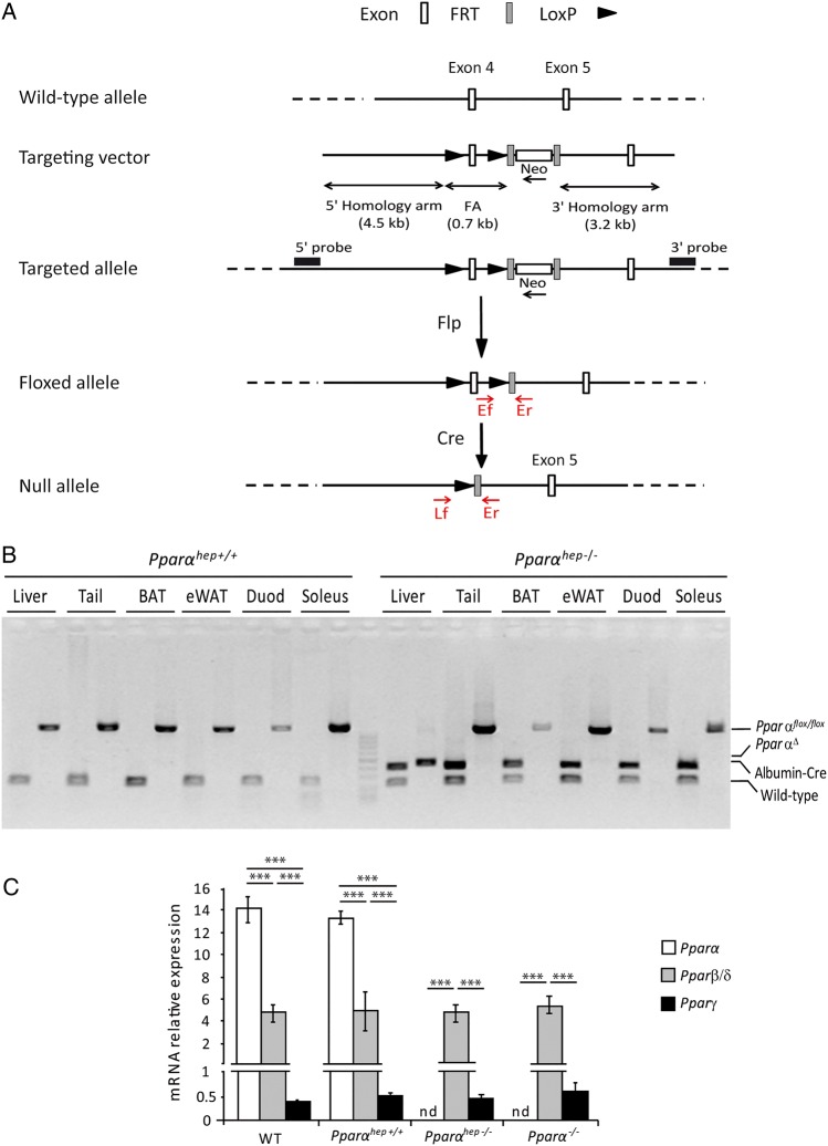
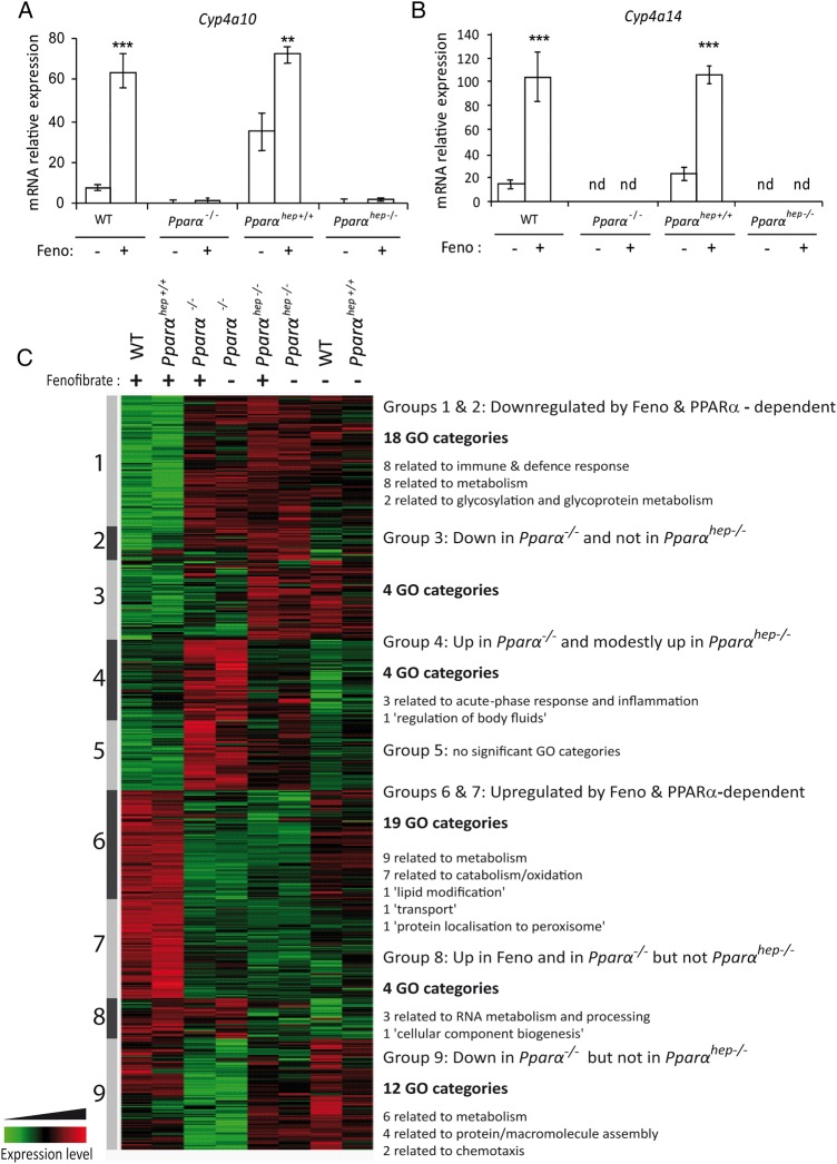
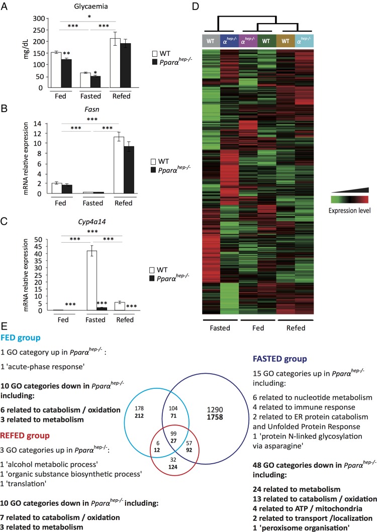
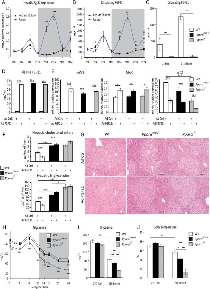
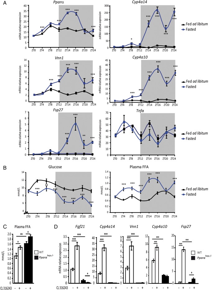
Design: We constructed a novel hepatocyte-specific PPARα knockout (Pparα(hep-/-)) mouse model. Using this novel model, we performed transcriptomic analysis following fenofibrate treatment. Next, we investigated which physiological challenges impact on PPARα. Moreover, we measured the contribution of hepatocytic PPARα activity to whole-body metabolism and fibroblast growth factor 21 production during fasting. Finally, we determined the influence of hepatocyte-specific PPARα deficiency in different models of steatosis and during ageing.

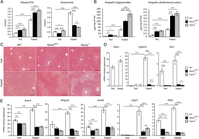
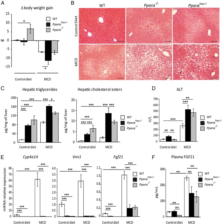
Results: Hepatocyte PPARα deletion impaired fatty acid catabolism, resulting in hepatic lipid accumulation during fasting and in two preclinical models of steatosis. Fasting mice showed acute PPARα-dependent hepatocyte activity during early night, with correspondingly increased circulating free fatty acids, which could be further stimulated by adipocyte lipolysis. Fasting led to mild hypoglycaemia and hypothermia in Pparα(hep-/-) mice when compared with Pparα(-/-) mice implying a role of PPARα activity in non-hepatic tissues. In agreement with this observation, Pparα(-/-) mice became overweight during ageing while Pparα(hep-/-) remained lean. However, like Pparα(-/-) mice, Pparα(hep-/-) fed a standard diet developed hepatic steatosis in ageing.

Conclusions: Altogether, these findings underscore the potential of hepatocyte PPARα as a drug target for NAFLD.

Keywords: GENE EXPRESSION; LIPID METABOLISM; NONALCOHOLIC STEATOHEPATITIS.


Figures

Figure 1
Figure 1
Characterisation of the hepatocyte-specific peroxisome proliferator-activated receptor α (PPARα) knockout mouse model. (A) Schematic of the targeting strategy to disrupt hepatic Pparα expression. (B) PCR analysis of Pparα floxed (Pparαhep+/+) and Albumin-Cre (Albumin-Cre+/−) genes from mice that are liver wild-type (WT), (Pparαhep+/+) or liver knockout (Pparαhep−/−) for Pparα using DNA extracted from different organs. (C) Relative mRNA expression levels of Pparα, Pparβ/δ and Pparγ from liver samples of WT, liver WT (Pparαhep+/+), Pparα liver knockout (Pparαhep−/−) and Pparα knockout (Pparα−/−) mice (n=8 mice per group). Data represent mean±SEM. ***p≤0.005. FA, floxed allele; Flp, flippase; FRT, flippase recognition target; LoxP, locus of X-overP1; nd, not detected; PparαΔ, Pparα deletion; WT, the Albumin-Cre−/− allele.
Figure 2
Figure 2
Pharmacological peroxisome proliferator-activated receptor α (PPARα) activation using fenofibrate reveals hepatocyte-specific PPARα-dependent biological functions. Liver samples from wild-type (WT), PPARα knockout (Pparα−/−), liver WT (Pparαhep+/+) and Pparα hepatocyte knockout (Pparαhep−/−) mice treated with fenofibrate (Feno, +) or vehicle (−) by oral gavage for 14 days were collected. (A and B) The relative gene expression of two specific PPARα target genes Cyp4a10 (A) and Cyp4a14 (B) was measured by qRT-PCR. Data represent mean±SEM. **p≤0.01, ***p≤0.005. (C) Heat map representing data from a microarray experiment performed with liver samples. Hierarchical clustering is also shown, which allows the definition of nine gene clusters. Gene Ontology (GO) analysis of each cluster revealed significant biological functions (p≤0.05). nd, not detected.
Figure 3
Figure 3
Hepatocyte-specific peroxisome proliferator-activated receptor α (PPARα) function is dependent on nutritional status. Wild-type (WT) and PPARα liver knockout (Pparαhep−/−) male 8-week-old mice were fed ad libitum, fasted for 24 h, or fasted for 24 h and refed for 24 h. All mice were killed at ZT14, and sera and livers were collected. (A) Quantification of circulating glucose levels. (B, C) Relative mRNA expressions of Fasn (B) and Cyp4a14 (C) in liver samples quantified by qRT-PCR. Data represent mean±SEM. *p≤0.05, **p≤0.01, ***p≤0.005. (D) Heat map was performed based on average gene expression levels from WT (n=12 (6 WT and 6 Pparαhep+/+)) and from Pparαhep−/− (n=6). (E) Venn diagram and associated Gene Ontology (GO) function analysis (p≤0.05), GO categories corresponding to functions down in the absence of PPARα are in bold, GO categories corresponding to functions up in the absence of PPARα are in regular font.
Figure 4
Figure 4
Fasting is the major inducer of hepatic peroxisome proliferator-activated receptor α (PPARα) activity. Wild-type (WT), hepatocyte-specific PPARα knockout (Pparαhep−/−) and total PPARα knockout (Pparα−/−) mice were fed ad libitum or fasted for 24 h and then killed. (A) Quantification of plasma free fatty acids (FFAs) and ketone bodies (ketonaemia). (B) Hepatic triglycerides and cholesterol esters hepatic levels. (C) Representative pictures of H&E staining of liver sections. Scale bars, 100 µm. (D) Relative mRNA expression levels of Pparα, Cyp4a14 and Vnn1 in liver samples determined by qRT-PCR. (E) Quantification of mRNA expression of Acox1, Hmgcs2, Acadl, Fsp27 and Plin5 by qRT-PCR. Data shown as mean±SEM. *p≤0.05, **p≤0.01, ***p≤0.005.
Figure 5
Figure 5
Hepatocyte and extrahepatocyte peroxisome proliferator-activated receptor α (PPARα) regulate fibroblast growth factor 21 (FGF21), glycaemia and body temperature during fasting. (A and B) Eleven-week-old male mice of the C57Bl/6J background were fed ad libitum or fasted for 24 h, and were killed around the clock from ZT0 to ZT24. (A) Fgf21 mRNA was quantified by qRT-PCR. (B) Quantification of circulating FGF21 levels by ELISA. (C) Twelve-week-old wild-type (WT), PPARα-hepatocyte knockout (Pparαhep−/−) and PPARα knockout (Pparα−/−) male mice were fed ad libitum or fasted for 16 h and blood was collected at ZT8 (ZT8 fed) or at ZT16 (ZT16 fasted). FGF21 plasma level was determined by ELISA. (D–G) Male mice of WT, Pparαhep−/− and Pparα−/− genotypes were infected with an adenoviral construct containing cDNA of Fgf21 or an empty vector. Mice were sacrificed after a 24 h fasting period at ZT14. (D) Quantification of circulating FGF21 levels by ELISA. (E) Fgf21, G6pd and Scd1 mRNAs were quantified by qRT-PCR. (F) Quantification of hepatic cholesterol esters and triglycerides. (G) Representative pictures of H&E staining of liver sections. Scale bars, 100 µm. (H) Plasma glucose level was monitored over a 24 h fasting period from ZT0 to ZT24 in WT, Pparαhep−/− and Pparα−/− mice. (I, J) Plasma glucose (I) and body temperature (J) were determined at ZT0 in fed mice or at ZT0 in mice fasted for 24 h. Data are shown as mean±SEM. *p≤0.05, **p≤0.01, ***p≤0.005.
Figure 6
Figure 6
Hepatocyte peroxisome proliferator-activated receptor α (PPARα) activity is induced by adipose tissue lipolysis. (A and B) Liver samples were collected from male wild-type (WT) C57Bl/6J mice that were fed ad libitum (black curve) or fasted (blue curve) over 24 h. (A) Hepatic mRNA expression levels of Pparα, Cyp4a14, Vnn1, Cyp4a10, Fsp27 and Tnfα were quantified by qRT-PCR. (B) Plasma glucose and free fatty acids (FFA) were measured. (C and D) WT and PPARα hepatocyte-specific knockout (Pparαhep−/−) mice were treated with the β3-adrenergic receptor agonist CL316243 at ZT6 and then killed at ZT14. (C) Quantification of plasma FFA. (D) Relative mRNA expression levels of Fgf21, Cyp4a14, Vnn1, Cyp4a10 and Fsp27 were measured by qRT-PCR. Data are shown as mean±SEM. *p≤0.05, **p≤0.01, ***p≤0.005.
Figure 7
Figure 7
Liver peroxisome proliferator-activated receptor α (PPARα) deficiency aggravates non-alcoholic steatohepatitis in response to a methionine-deficient and choline-deficient diet (MCD). Wild-type (WT), PPARα hepatocyte knockout (Pparαhep−/−) and PPARα knockout (Pparα−/−) mice were fed a MCD or a control diet for 2 weeks and were killed at ZT8. (A) Body weight gain was measured over 2 weeks. (B) Representative pictures of H&E staining on liver sections. Scale bar, 100 µm. (C) Quantification of hepatic triglycerides and cholesterol esters. (D) Alanine transaminase activity level in plasma. (E) Hepatic mRNA expression levels of Cyp4a14, Vnn1 and Fgf21. (F) Plasma levels of fibroblast growth factor 21 (FGF21). Data are shown as mean±SEM. *p≤0.05, **p≤0.01, ***p≤0.005.
Figure 8
Figure 8
Mice deficient in hepatic peroxisome proliferator-activated receptor α (PPARα) develop spontaneous hepatic steatosis during ageing. Wild-type (WT), PPARα hepatocyte knockout (Pparαhep−/−) and PPARα knockout (Pparα−/−) mice were fed a chow diet for 51 weeks. All mice were killed at ZT16 in a non-fasted state. (A) Body weight gain was followed over time. (B) Comparison of body weight between weeks 11 and 50. (C) Representative pictures of 52-week-old mice of the three genotypes. (D) Representative images of H&E staining of liver sections. Scale bar, 100 µm. (E) Quantification of hepatic triglycerides and cholesterol esters. (F) Measurement of plasma total cholesterol, HDL cholesterol and LDL cholesterol. (G) Fasting glycaemia. Data are shown as mean±SEM. *p≤0.05, **p≤0.01, ***p≤0.005.
Figure 9
Figure 9
Overview of hepatocyte-specific peroxisome proliferator-activated receptor α (PPARα)-regulated genes involved in fatty acid metabolism. This figure was designed based on transcriptome analysis of PPARα-dependent gene expression in hepatocytes. Genes listed in regular font are induced by fenofibrate and by fasting in wild-type (WT) but not in Pparαhep−/− mice. Genes in italics are repressed by fenofibrate and by fasting in WT but not in Pparαhep−/− mice. Genes referenced in bold are downregulated in Pparαhep−/− compared with WT mice, whatever the conditions.

Comment in

References

    1. Browning JD, Horton JD. Molecular mediators of hepatic steatosis and liver injury. J Clin Invest 2004;114:147–52. 10.1172/JCI22422 - DOI - PMC - PubMed
    1. Wahli W, Michalik L. PPARs at the crossroads of lipid signaling and inflammation. Trends Endocrinol Metab 2012;23:351–63. 10.1016/j.tem.2012.05.001 - DOI - PubMed
    1. Braissant O, Foufelle F, Scotto C, et al. . Differential expression of peroxisome proliferator-activated receptors (PPARs): tissue distribution of PPAR-alpha, -beta, and -gamma in the adult rat. Endocrinology 1996;137:354–66. 10.1210/endo.137.1.8536636 - DOI - PubMed
    1. Postic C, Girard J. Contribution of de novo fatty acid synthesis to hepatic steatosis and insulin resistance: lessons from genetically engineered mice. J Clin Invest 2008;118:829–38. 10.1172/JCI34275 - DOI - PMC - PubMed
    1. Kersten S. Integrated physiology and systems biology of PPARα. Mol Metab 2014;3:354–71. 10.1016/j.molmet.2014.02.002 - DOI - PMC - PubMed

MeSH terms