. 2016 May;142(5):981-93.
doi: 10.1007/s00432-016-2117-4. Epub 2016 Feb 2.

Evaluation of role of Notch3 signaling pathway in human lung cancer cells

Affiliations

Evaluation of role of Notch3 signaling pathway in human lung cancer cells

Wael Abdo Hassan et al. J Cancer Res Clin Oncol. 2016 May.

Abstract

There is still a debate on the extent to which Notch3 signaling is involved in lung carcinogenesis and whether such function is dependent on cancer type or not.

Purpose: To evaluate Notch3 expression in different types of human lung cancer cells.

Methods: Notch3 was detected in human lung cancer cell lines and in tissues. Then, small interfering RNA (siRNA) was used to down-regulate the expression of Notch3 in H69AR small cell lung carcinoma (SCLC) cells; two non-small cell lung carcinoma (NSCLC) cells; A549 adenocarcinoma (ADC); and H2170 squamous cell carcinoma (SCC). In addition, Notch3 intracellular domain (N3ICD) plasmid was transfected into H1688 human SCLC cells. We observed the effect of deregulating Notch3 signaling on the following cell properties: Notch-related proteins, cell morphology, adhesion, epithelial-mesenchymal transition (EMT), motility, proliferation and neuroendocrine (NE) features of SCLC.

Results: Notch3 is mainly expressed in NSCLC, and the expression of Notch1, Hes1 and Jagged1 is affected by Notch3. Notch3 has opposite functions in SCLC and NSCLC, being a tumor suppressor in the former and tumor promoting in the latter, in the context of cell adhesion, EMT and motility. Regarding cell proliferation, we found that inhibiting Notch3 in NSCLC decreases cell proliferation and induces apoptosis in NSCLC. Notch3 has no effect on cell proliferation or NE features of SCLC.

Conclusion: Notch3 signaling in lung carcinoma is dependent on cell type. In SCLC, Notch3 behaves as a tumor suppressor pathway, while in NSCLC it acts as a tumor-promoting pathway.

Keywords: Human lung cancer; Notch3 intracellular domain (N3ICD) plasmid; Notch3 signaling; Small interfering RNA (siRNA).


Conflict of interest statement

The authors declare that they have no conflict of interest.

Figures

Fig. 1
Fig. 1
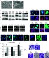
Expression of Notch3 in human lung cancer, conforming knocking down (KD) and overexpression of Notch3, and examining its effect on Notch-related proteins (c-Myc, Hes1, Jagged1 and Jagged2). a Notch3 protein expression in human small cell lung cancer (SCLC); non-small cell lung cancer (NSCLC) cell lines; adenocarcinoma (ADC); and squamous cell carcinoma (SCC). Cells cultured in the same conditions were harvested and supplied for western blotting (WB). The expression of β-actin was used as an internal control. Notch3 [full length and intracellular domain (ICD)] was mainly detected in NSCLC cells, and only in H69AR and SBC-3 SCLC cells. The experiment was performed in triplicate. b Immunofluorescence (IFA) staining of Notch3 in selected lung cancer cells. The signal of the molecule (Alexa Fluors 488, green) was clearly detectable in the cytoplasm of H69AR, A549 and H2170 cells, while it was absent in H1688 cells. Nuclei were counterstained with 4′, 6-diamidino-2-phenylindole (DAPI). Original magnification, ×400. c Representative photographs of immunohistochemical (IHC) staining of Notch3 in different lung cancer tissues. Tissues were immunohistochemically stained using an antibody against Notch3. The cytoplasmic and membranous immunoreactions (brown) for Notch3 were evident in NSCLC cells, especially ADC, while it was absent in SCLC cells. Original magnification, ×400. Scale bar 20 µm. d WB of Notch3 and related proteins (Notch1, Notch2, Hes1, Hey1, Jagged1, Jagged2, c-Myc, Akt and pAkt) 48 h after transfection. Note the decreased expressions of Notch3, Notch1, Hes1 and Jagged1 in the Notch3 siRNA-treated cells and their increased expression (except for Notch1) in H1688 cells transfected with N3ICD plasmid. In addition, Jagged2 expression was decreased in Notch3 siRNA-treated H69AR and A549 cells. In addition, c-Myc expression was decreased in Notch3 siRNA-treated A549 cells. No change in the expression level of Notch2, Hey1, Akt and pAKt proteins. The expression of β-actin was used as an internal control. The experiment was performed in triplicate. Hes, hairy and enhancer of split; Hey, hairy/enhancer of split related to YRPW motif; pAKT, phosphorylated Akt. e Comparative quantitation of Hes1 and c-Myc proteins in H1688 cells transfected with N3ICD compared to control cells was performed by an ATTO CS analyzer, and the ratios relative to β-actin protein were determined. Note the increased expression of Hes1 protein in cells transfected with N3ICD plasmid, while no significant change in the expression of c-Myc protein was observed. The results represent the average of three independent experiments
Fig. 1
Fig. 1
Expression of Notch3 in human lung cancer, conforming knocking down (KD) and overexpression of Notch3, and examining its effect on Notch-related proteins (c-Myc, Hes1, Jagged1 and Jagged2). a Notch3 protein expression in human small cell lung cancer (SCLC); non-small cell lung cancer (NSCLC) cell lines; adenocarcinoma (ADC); and squamous cell carcinoma (SCC). Cells cultured in the same conditions were harvested and supplied for western blotting (WB). The expression of β-actin was used as an internal control. Notch3 [full length and intracellular domain (ICD)] was mainly detected in NSCLC cells, and only in H69AR and SBC-3 SCLC cells. The experiment was performed in triplicate. b Immunofluorescence (IFA) staining of Notch3 in selected lung cancer cells. The signal of the molecule (Alexa Fluors 488, green) was clearly detectable in the cytoplasm of H69AR, A549 and H2170 cells, while it was absent in H1688 cells. Nuclei were counterstained with 4′, 6-diamidino-2-phenylindole (DAPI). Original magnification, ×400. c Representative photographs of immunohistochemical (IHC) staining of Notch3 in different lung cancer tissues. Tissues were immunohistochemically stained using an antibody against Notch3. The cytoplasmic and membranous immunoreactions (brown) for Notch3 were evident in NSCLC cells, especially ADC, while it was absent in SCLC cells. Original magnification, ×400. Scale bar 20 µm. d WB of Notch3 and related proteins (Notch1, Notch2, Hes1, Hey1, Jagged1, Jagged2, c-Myc, Akt and pAkt) 48 h after transfection. Note the decreased expressions of Notch3, Notch1, Hes1 and Jagged1 in the Notch3 siRNA-treated cells and their increased expression (except for Notch1) in H1688 cells transfected with N3ICD plasmid. In addition, Jagged2 expression was decreased in Notch3 siRNA-treated H69AR and A549 cells. In addition, c-Myc expression was decreased in Notch3 siRNA-treated A549 cells. No change in the expression level of Notch2, Hey1, Akt and pAKt proteins. The expression of β-actin was used as an internal control. The experiment was performed in triplicate. Hes, hairy and enhancer of split; Hey, hairy/enhancer of split related to YRPW motif; pAKT, phosphorylated Akt. e Comparative quantitation of Hes1 and c-Myc proteins in H1688 cells transfected with N3ICD compared to control cells was performed by an ATTO CS analyzer, and the ratios relative to β-actin protein were determined. Note the increased expression of Hes1 protein in cells transfected with N3ICD plasmid, while no significant change in the expression of c-Myc protein was observed. The results represent the average of three independent experiments
Fig. 2
Fig. 2
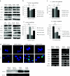
Effect of Notch3 on cell morphology, adhesion, epithelial-mesenchymal transition (EMT) markers (E cadherin, snail, slug, vimentin and twist), cell motility and invasion. a Images of H69AR cells transfected with Notch3 siRNA, through light microscope. Note the loss of cell-to-cell adhesion (arrows). Original magnification, ×200. b WB and IFA analysis of EMT markers, 48 h after transfection. Note the decreased E cadherin (E cad) and the increased snail, slug and vimentin (Vim) expressions in H69AR cells transfected with Notch3 siRNA. On the other hand, note the increased E cad and the decreased Vim expressions in H1688 cells with N3ICD, as well as in NSCLC cells transfected with Notch3 siRNA, in addition to decreased snail expression in the latter. The expression of β-actin was used as an internal control. The experiment was performed in triplicate. The IFA photographs represent staining for E cad and Vim. Each protein was detected using specific antibodies. The changes in the signal of E cad (Alexa Fluors 488, green) and in the signal of Vim (Alexa Fluors 568, red) were clearly detected in the membrane and cytoplasm of cells. Original magnification, ×400. c IFA staining for gamma laminin 2 chain alpha (GL2), a cell motility marker. The changes in the signal of GL2 (Alexa Fluors 488, green) were clearly detected in the cytoplasm of cells. Inhibiting Notch 3 in H69AR cells increased cytoplasmic expression of GL2, while in H1688 with N3ICD and in NSCLC cells transfected with Notch3 siRNA, its expression decreased. Original magnification, ×400. d In vitro Matrigel transmembrane invasion assay. Cells were incubated with either control or Notch3-specific siRNA for 24 h. Cells that penetrated the Matrigel-coated membrane were fixed and stained. The numbers of invaded cells obtained from three independent experiments were counted and statistically analyzed. The bar graphs represent the average numbers of cells on the underside of the membranes. The Y-axis shows the numbers of cells invading the underside of the membranes. The photographs are representative fields of invasive cells on the membranes. Original magnification, ×100. Inhibiting Notch3 in H69AR cells increased the number of invading cells, while its inhibition in NSCLC cells decreased number of invading cells. Mean ± S.D. of three independent experiments. p value analyzed by Student’s t-test. GL2, gamma laminin 2 chain alpha protein
Fig. 3
Fig. 3
Effect of Notch3 on cell proliferation, apoptosis and neuroendocrine (NE) differentiation of lung cancer cells. a WB analysis for cell proliferation and apoptotic markers in cells, 48 h after transfection. Note the increased expression of phosphorylated histone H3 (pH3) and caspase 3 (Csp3) proteins both in H69AR cells transfected with Notch3 siRNA and in H1688 cells with N3ICD, with weak induction of cleaved caspase 3 (CCsp3) in the later. In NSCLC, note the decreased pH3 and the increased Csp3 and CCsp3 proteins in cells transfected with Notch3 siRNA. No changes in the expression levels of Bcl-2 and pBcl-2. The expression of β-actin was used as an internal control. The experiment was performed in triplicate. b In situ apoptotic assay. The bar graph represents the average numbers of green-emitting apoptotic cells, counted 48 h after transfection, plotted on Y-axis. Apoptotic cells with fragmented DNA typically emit green fluorescein from the incorporation of terminal deoxynucleotidyl transferase to label 3′-OH ends of DNA. There was no statistical significant difference in either H69AR cells transfected with Notch3 siRNA or H1688 cells with N3ICD, when compared to their subsequent controls. However, in NSCLC cells transfected with Notch3 siRNA, the number of cells emitting green apoptotic signals was increased, when compared to their subsequent controls. Mean ± S.D. of three independent experiments. p value analyzed by Student’s t-test. c Cell counting assay. The bar graph represents the average numbers of cells (×104), counted 48 h after transfection, using the described cell counting assay, plotted on Y-axis. There was no statistically significant difference in either H69AR cells transfected with Notch3 siRNA or H1688 cells with N3ICD, when compared to their subsequent controls. However, in NSCLC cells transfected with Notch3 siRNA, the reduction in number of cells was statistically significant, when compared to their subsequent controls. Mean ± S.D. of three independent experiments. p value analyzed by Student’s t-test. pH3, phosphorylated histone H3; pBcl2, phosphorylated Bcl2; Csp3, caspase 3; CCsp3, cleaved caspase 3. d IFA, WB and RT-PCR analysis of NE markers, 48 h after transfection. The IFA photographs represent staining of ASCL-1 and CGA. NE markers. The signals of ASCL-1 (Alexa Fluors 568, red) and of CGA (Alexa Fluors 488, green) were detected in nuclei (DAPI, blue) and cytoplasm of H1688 cells, respectively. No change in the expression level of NE markers was noted. Original magnification, ×400. WB analysis for ASCL-1 protein expression in SCLC cells used. No change in its expression, regardless of Notch3 expression. The expression of β-actin was used as an internal control. The experiment was performed in triplicate. RT-PCR shows the level of mRNA expressions of Notch3 and NE markers in cancer cells, 48 h after transfection. Notch3 mRNA level expression was decreased in H69AR cells transfected with Notch3 siRNA, while increased in H1688 cells transfected with N3ICD. No significant change in the expression level of NE markers was noted. The expression of β-actin was used as an internal control. The experiment was performed in triplicate. ASCL, achaete-scute homolog; CGA, chromogranin-A; SYP, synaptophysin; CGRP, calcitonin gene-related peptide; NSE, neuron-specific enolase; GRP, gastrin-releasing peptide
Fig. 4
Fig. 4
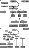
Molecular pathway for Notch3 signaling in (A) human SCLC and (B) human NSCLC. a Effect of Notch3 expression in SCLC. H69AR and H1688 cells are representatives of SCLC. Jagged1 is regulated by Notch3 in SCLC cells. Hes1 expression is affected by Notch3 in SCLC cells. In addition, the expressions of Jagged2 and thus Notch1 are affected by Notch3 expression in H69AR cells. Notch3 expression has no statistically significant effect on cell proliferation or NE features of SCLC cells. However, its expression inhibits EMT molecules, as twist and Vim in both H69AR and H1688 cells and snail and slug in H69AR, leading to increased E cad expression and decreased GL2 expression, resulting in increased cell adhesion and decreased cell motility and invasion. b Effect of Notch3 expression in NSCLC. A549 and H2170 cells are representatives of NSCLC. Jagged1 is regulated by Notch3 in NSCLC cells. In addition, the expression of Notch1 is affected by Notch3. Moreover, in A549 cells, the expression of Jagged2 and c-Myc are affected by Notch3. Expression of Notch3 increases the expression of EMT molecules, as snail and vim in both A549 and H2170 cells, in addition to twist in H2170, leading to decreased E cad expression and increased GL2 expression, resulting in decreased cell adhesion and increased cell motility and invasion. In addition, expression of Notch3 increases the expression of cell proliferation marker, p-H3, and inhibits the expression of apoptotic marker, CCsp3, resulting in increased cell proliferation and inhibition of cell apoptosis. E cad, E cadherin; Vim, vimentin; GL2, gamma laminin 2 chain alpha protein; p-H3, phosphorylated histone H3; CCsp3, cleaved caspase 3

References

    1. Arasada RR, Amann JM, Rahman MA, Huppert SS, Carbone DP (2014) EGFR blockade enriches for lung cancer stem-like cells through Notch3-dependent signaling. Cancer Res 74(19):5572–5584 - PMC - PubMed
    1. Bunn PA Jr (2012) Worldwide overview of the current status of lung cancer diagnosis and treatment. Arch Pathol Lab Med 136:1478–1481 - PubMed
    1. Chen X, Stoeck A, Lee SJ, IeM Shih, Wang MM, Wang TL (2010) Jagged1 expression regulated by Notch3 and Wnt/β-catenin signaling pathways in ovarian cancer. Oncotarget 3:210–218 - PMC - PubMed
    1. Collins BJ, Kleeberger W, Ball DW (2004) Notch in lung development and lung cancer. Semin Cancer Biol 14(5):357–364 - PubMed
    1. Dang TP, Gazdar AF, Virmani AK, Sepetavec T, Hande KR, Minna JD, Roberts JR, Carbone DP (2000) Chromosome 19 translocation, overexpression of Notch3, and human lung cancer. J Natl Cancer Inst 92:1355–1357 - PubMed

Publication types

MeSH terms