

Culturing on decellularized extracellular matrix enhances antioxidant properties of human umbilical cord-derived mesenchymal stem cells
- PMID: 26838870
- PMCID: PMC9805354
- DOI: 10.1016/j.msec.2015.12.090
Culturing on decellularized extracellular matrix enhances antioxidant properties of human umbilical cord-derived mesenchymal stem cells
Abstract
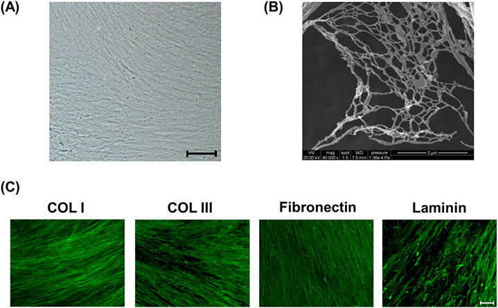
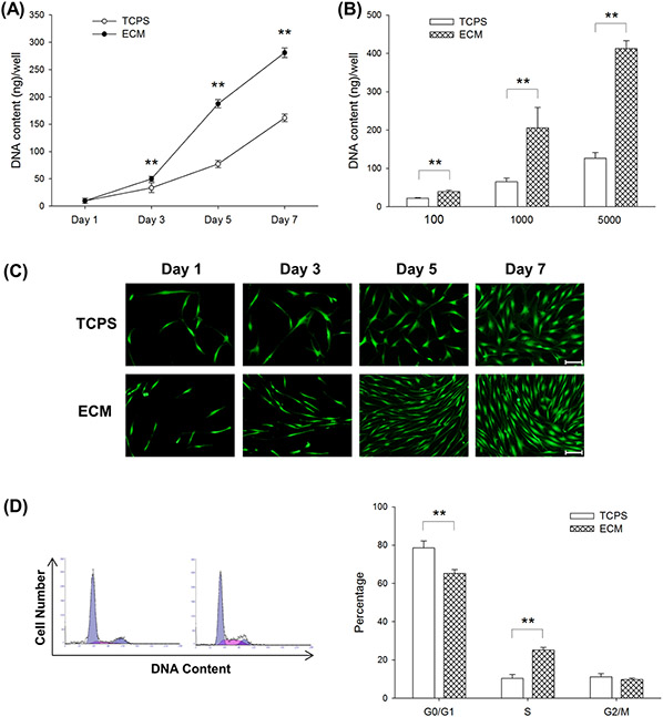
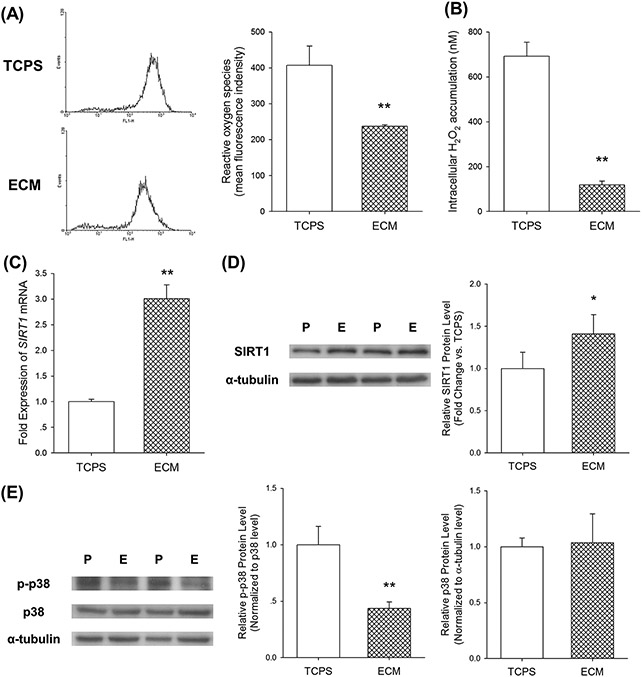
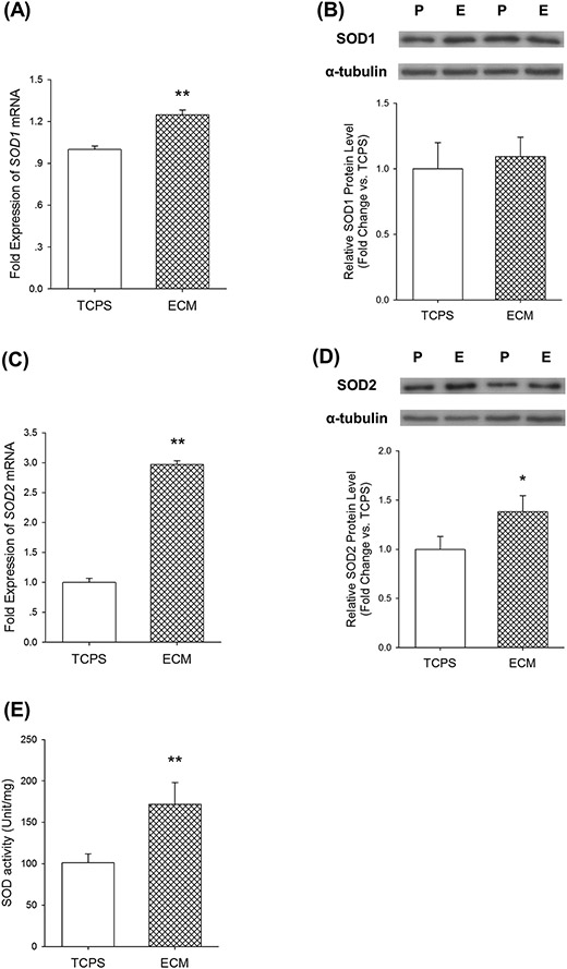
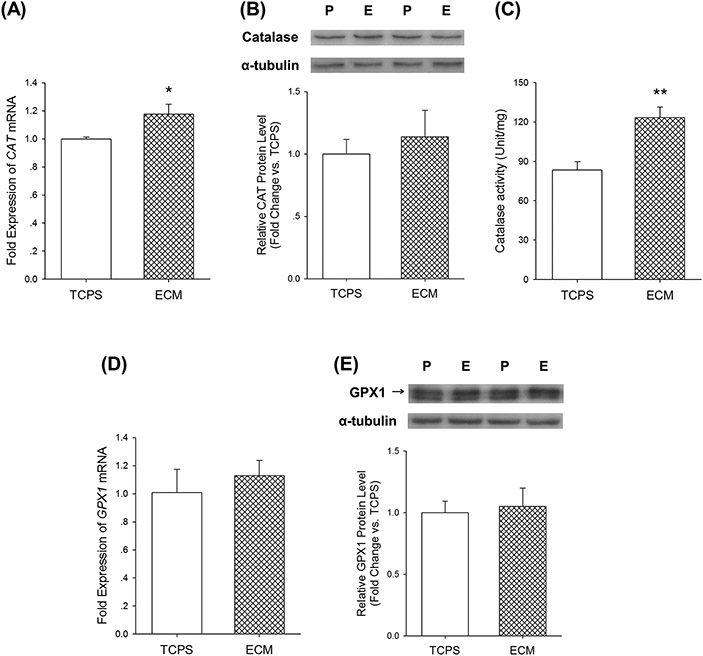
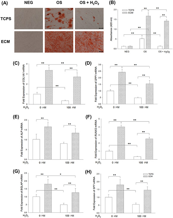
Human umbilical cord-derived mesenchymal stem cells (UC-MSCs) have attracted great interest in clinical application because of their regenerative potential and their lack of ethical issues. Our previous studies showed that decellularized cell-deposited extracellular matrix (ECM) provided an in vivo-mimicking microenvironment for MSCs and facilitated in vitro cell expansion. This study was conducted to analyze the cellular response of UC-MSCs when culturing on the ECM, including reactive oxygen species (ROS), intracellular antioxidative enzymes, and the resistance to exogenous oxidative stress. After decellularization, the architecture of cell-deposited ECM was characterized as nanofibrous, collagen fibrils and the matrix components were identified as type I and III collagens, fibronectin, and laminin. Compared to tissue culture polystyrene (TCPS) plates, culturing on ECM yielded a 2-fold increase of UC-MSC proliferation and improved the percentage of cells in the S phase by 2.4-fold. The levels of intracellular ROS and hydrogen peroxide (H2O2) in ECM-cultured cells were reduced by 41.7% and 82.9%, respectively. More importantly, ECM-cultured UC-MSCs showed enhanced expression and activity of intracellular antioxidative enzymes such as superoxide dismutase and catalase, up-regulated expression of silent information regulator type 1, and suppressed phosphorylation of p38 mitogen-activated protein kinase. Furthermore, a continuous treatment with exogenous 100μM H2O2 dramatically inhibited osteogenic differentiation of UC-MSCs cultured on TCPS, but culturing on ECM retained the differentiation capacity for matrix mineralization and osteoblast-specific marker gene expression. Collectively, by providing sufficient cell amounts and enhancing antioxidant capacity, decellularized ECM can be a promising cell culture platform for in vitro expansion of UC-MSCs.
Keywords: Antioxidative enzymes; Extracellular matrix; Mesenchymal stem cells; Osteogenesis; Reactive oxygen species.
Copyright © 2015 Elsevier B.V. All rights reserved.
Conflict of interest statement
Conflicts of interest
The authors declare no conflicts of interest.
Figures
Similar articles
-
SIRT1-dependent anti-senescence effects of cell-deposited matrix on human umbilical cord mesenchymal stem cells.J Tissue Eng Regen Med. 2018 Feb;12(2):e1008-e1021. doi: 10.1002/term.2422. Epub 2017 Jun 20. J Tissue Eng Regen Med. 2018. PMID: 28107614 Free PMC article.
-
Inhibition of osteoclastogenesis by stem cell-derived extracellular matrix through modulation of intracellular reactive oxygen species.Acta Biomater. 2018 Apr 15;71:118-131. doi: 10.1016/j.actbio.2018.03.003. Epub 2018 Mar 8. Acta Biomater. 2018. PMID: 29526830 Free PMC article.
-
Extracellular matrix modulates the biological effects of melatonin in mesenchymal stem cells.J Endocrinol. 2014 Nov;223(2):167-80. doi: 10.1530/JOE-14-0430. Epub 2014 Sep 10. J Endocrinol. 2014. PMID: 25210047
-
Native and solubilized decellularized extracellular matrix: A critical assessment of their potential for improving the expansion of mesenchymal stem cells.Acta Biomater. 2017 Jun;55:1-12. doi: 10.1016/j.actbio.2017.04.014. Epub 2017 Apr 12. Acta Biomater. 2017. PMID: 28412553 Review.
-
Age associated communication between cells and matrix: a potential impact on stem cell-based tissue regeneration strategies.Organogenesis. 2014;10(3):289-98. doi: 10.4161/15476278.2014.970089. Organogenesis. 2014. PMID: 25482504 Free PMC article. Review.
Cited by
-
A prospect of cell immortalization combined with matrix microenvironmental optimization strategy for tissue engineering and regeneration.Cell Biosci. 2019 Jan 5;9:7. doi: 10.1186/s13578-018-0264-9. eCollection 2019. Cell Biosci. 2019. PMID: 30627420 Free PMC article. Review.
-
Extracellular matrix deposited by Wharton's jelly mesenchymal stem cells enhances cell expansion and tissue specific lineage potential.Am J Transl Res. 2018 Nov 15;10(11):3465-3480. eCollection 2018. Am J Transl Res. 2018. PMID: 30662600 Free PMC article.
-
Hypoxic Extracellular Matrix Preserves Its Competence after Expansion of Human MSCs under Physiological Hypoxia In Vitro.Biomimetics (Basel). 2023 Oct 7;8(6):476. doi: 10.3390/biomimetics8060476. Biomimetics (Basel). 2023. PMID: 37887607 Free PMC article.
-
Cell-Derived Extracellular Matrix for Tissue Engineering and Regenerative Medicine.Front Bioeng Biotechnol. 2020 Dec 3;8:602009. doi: 10.3389/fbioe.2020.602009. eCollection 2020. Front Bioeng Biotechnol. 2020. PMID: 33344434 Free PMC article. Review.
-
In vitro comparison of harvesting site effects on cardiac extracellular matrix hydrogels.J Biomed Mater Res A. 2021 Oct;109(10):1922-1930. doi: 10.1002/jbm.a.37184. Epub 2021 Apr 6. J Biomed Mater Res A. 2021. PMID: 33822464 Free PMC article.
References
-
- Pittenger MF, et al., Multilineage potential of adult human mesenchymal stem cells, Science 284 (5411) (1999) 143–147. - PubMed
-
- Barry F, Murphy M, Mesenchymal stem cells in joint disease and repair, Nat. Rev. Rheumatol 9 (10) (2013) 584–594. - PubMed
-
- Pappa KI, Anagnou NP, Novel sources of fetal stem cells: where do they fit on the developmental continuum?, Regen. Med 4 (3) (2009) 423–433. - PubMed
-
- Corrao S, et al., New frontiers in regenerative medicine in cardiology: the potential of Wharton's jelly mesenchymal stem cells, Curr. Stem Cell Res. Ther 8 (1) (2013) 39–45. - PubMed
Publication types
MeSH terms
Substances
Grants and funding
LinkOut - more resources
Full Text Sources
Other Literature Sources
Medical
