. 2016 Mar 15;133(11):1093-103.
doi: 10.1161/CIRCULATIONAHA.115.020918. Epub 2016 Feb 2.

Endothelial p110γPI3K Mediates Endothelial Regeneration and Vascular Repair After Inflammatory Vascular Injury

Affiliations

Endothelial p110γPI3K Mediates Endothelial Regeneration and Vascular Repair After Inflammatory Vascular Injury

Xiaojia Huang et al. Circulation. .

Abstract

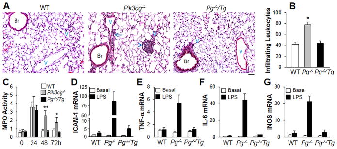
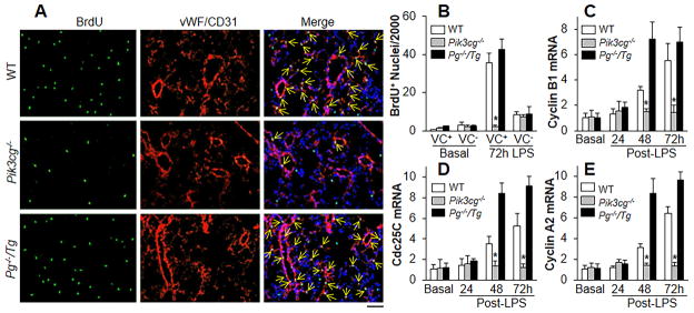
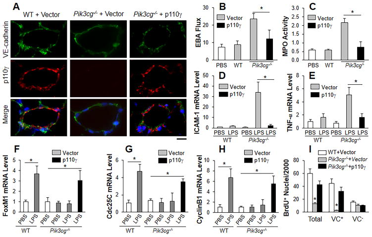
Background: The integrity of endothelial monolayer is a sine qua non for vascular homeostasis and maintenance of tissue-fluid balance. However, little is known about the signaling pathways regulating regeneration of the endothelial barrier after inflammatory vascular injury.

Methods and results: Using genetic and pharmacological approaches, we demonstrated that endothelial regeneration selectively requires activation of p110γPI3K signaling, which thereby mediates the expression of the endothelial reparative transcription factor Forkhead box M1 (FoxM1). We observed that FoxM1 induction in the pulmonary vasculature was inhibited in mice treated with a p110γ-selective inhibitor and in Pik3cg(-/-) mice after lipopolysaccharide challenge. Pik3cg(-/-) mice exhibited persistent lung inflammation induced by sepsis and sustained increase in vascular permeability. Restoration of expression of either p110γ or FoxM1 in pulmonary endothelial cells of Pik3cg(-/-) mice restored endothelial regeneration and normalized the defective vascular repair program. We also observed diminished expression of p110γ in pulmonary vascular endothelial cells of patients with acute respiratory distress syndrome, suggesting that impaired p110γ-FoxM1 vascular repair signaling pathway is a critical factor in persistent leaky lung microvessels and edema formation in the disease.

Conclusions: We identify p110γ as the critical mediator of endothelial regeneration and vascular repair after sepsis-induced inflammatory injury. Thus, activation of p110γ-FoxM1 endothelial regeneration may represent a novel strategy for the treatment of inflammatory vascular diseases.

Keywords: endothelial regeneration; endothelium, vascular; inflammation; vascular diseases; vascular repair.


Figures

Figure 1
Figure 1
p110γ mediates FoxM1 expression during the repair phase following LPS challenge. (A) FoxM1 mRNA expression in lungs. At 12h post-LPS, WT mice were administered either DMSO (CTL) or wortmannin (Wor, 0.05mg/kg BW, i.p.) every 12 h. Lung tissue was collected for QRT-PCR analysis. n = 5 mice/group. *, P < 0.05; **, P < 0.01 (Student t test). (B) Representative Western blotting demonstrating decreased FoxM1 protein expression induced by wortmannin treatment. The experiment was repeated twice with similar results. (C) Inhibition of p110γ but not p110α decreased FoxM1 expression. At 12 h post-LPS, WT mice were administered either DMSO (CTL), the p110γ inhibitor AS-605240 (AS) (30 mg/kg, per os.), or the p110α inhibitor PI-103 (10 mg/kg, i.p.) at 12 h intervals. Lungs at 72h post-LPS were collected for QRT-PCR analysis. n = 5. *, P < 0.05 (ANOVA). (D) Inhibition of FoxM1 expression in Pik3cg−/− mouse lungs following LPS challenge. n = 5. *, P < 0.05; **, P < 0.01 (Student’s t test).
Figure 2
Figure 2
Impairment of vascular repair in Pik3cg−/− mice secondary to inhibition of FoxM1 expression. (A) Pulmonary transvascular EBA flux demonstrating defective vascular repair in Pik3cg−/− mouse lungs which was rescued by overexpression of FoxM1 in Pg−/−/Tg mice. n = 5. *, P < 0.05; *, P < 0.01 (ANOVA). (B) Scanning electron microscopy demonstrating p110γ-mediated FoxM1 expression is required for vascular repair. Representative micrographs of lung sections from mice challenged with LPS for 60h are shown. Pik3cg−/− mouse lungs had extensive extravasation (Ex) of methacrylate tracer on the cut surfaces and many alveoli were filled with the tracer. However, Pg−/−/Tg lungs displayed normal profile similar to WT lungs. A, airways. The experiment was performed 3 times with similar data. Scale bar, 20μm. (C) Representative Western blotting of p110γ and FoxM1 in lung lysates at basal. Experiment were performed three times with similar results. (D) Representative micrographs of cremaster muscle venule demonstrating marked leakage of FITC-conjugated dextran in Pik3cg−/− mice in contrast to WT and Pg−/−/Tg at 48h post-LPS. Thirty min post-administration of FITC-conjugated dextran (i.v.), vascular permeability in the cremaster muscle venule was monitored by the FITC signal in an area of 0.02 mm2. The vessel walls were indicated in white lines. Scale bar, 10μm. (E) Graphic presentation of prominent vascular leakiness in Pik3cg−/− mice at 48h post-LPS challenge, which was rescued by FoxM1 overexpression in Pg−/−/Tg mice. n = 6 venules in 3 mice/group. *, P < 0.0001 (ANOVA).
Figure 3
Figure 3
Restoration of FoxM1 expression in Pg−/−/Tg mice mitigates lung inflammation. (A) H & E staining of lung sections (of 3 independent experiments) showing perivascular leukocyte infiltration in Pik3cg−/− mouse lungs at 48h post-LPS challenge. Arrows indicate leukocyte infiltration. Scale bar, 50 μm. Br, bronchia; V, vessel. (B) Analysis of infiltrating leukocytes in lungs at 48h post-LPS challenge. Bar graphs show infiltrating leukocytes per vessels (> 30μm in diameter). n = 5. *, P < 0.05(ANOVA). P−/−, Pik3cg−/−. (C) Time course of MPO activity in mouse lungs following LPS challenge (7.5 mg/kg, i.p.). n =5. *, P < 0.05; **, P < 0.001 (ANOVA). (D–G) QRT-PCR analysis of expression of proinflammatory mediators in mouse lungs. AT 48h post-LPS challenge, mouse lungs were collected for QRT-PCR analysis. n = 5. Elevated expression of pro-inflammatory mediators seen in Pik3cg−/− mouse lungs was inhibited in Pg−/−/Tg mouse lungs.
Figure 4
Figure 4
Impairment in endothelial cell proliferation in Pik3cg−/− lungs is rescued by expression of FoxM1. (A) Representative micrographs showing EC proliferation. Cryosections of lungs (3–5μm thick), collected at 72h following LPS challenge, were stained with FITC-conjugated anti-BrdU antibody to identify proliferating cells (green) and with anti-vWF and anti-CD31 antibodies to identify EC (red). Nuclei were counterstained with DAPI (blue). Arrows indicate proliferating lung ECs. Scale bar, 50 μm. (B) Graphic presentation of decreased proliferating ECs in Pik3cg−/− lungs. Three consecutive cryosections from each mouse lung were examined and average number of BrdU-positive nuclei was used. n = 5. *, P < 0.0001 (ANOVA). VC+, vWF+/CD31+ cells. (CE) QRT-PCR analysis of expression of FoxM1 target genes essential for cell cycle progression. n = 5, *, P < 0.01 (ANOVA).
Figure 5
Figure 5
Restored expression of p110γ in lung endothelial cells of Pik3cg−/− mice induces FoxM1 expression and normalizes endothelial regeneration. (A) Representative micrographs showing endothelial expression of p110γ in pulmonary vascular ECs of Pik3cg−/− mice at 30h following liposome-mediated transduction of p110γ plasmid DNA (driven by human CDH5 promoter). Scale bar, 30μm. (B) Endothelial expression of p110γ normalized the defective vascular repair phenotype of Pik3cg−/− mouse lungs. At 30h post-plasmid DNA transduction, mice were challenged with LPS and lungs were collected for assessing pulmonary transvascular EBA flux at 60h post-LPS. n = 5. *, P < 0.01 (t test). (C) Lung tissue MPO activity indicating resolved lung inflammation in Pik3cg−/− mice transduced with p110γ plasmid DNA at 60h post-LPS. *, P < 0.01(t test). (D, E) QRT-PCR analysis of expression of pro-inflammatory molecules in mouse lungs. *, P < 0.01 (t test). (FH) QRT-PCR analysis of FoxM1 expression and its downstream target genes in Pik3cg−/− lungs transduced with p110γ plasmid DNA. *, P < 0.01 (t test). (I) Endothelial expression of p110γ normalized lung EC proliferation in Pik3cg−/− mice at 60h post-LPS. *, P < 0.001 (ANOVA). VC, vWF/CD31.
Figure 6
Figure 6
Endothelial expression of FoxM1 rescued the defective vascular repair phenotype of Pik3cg−/− mouse lungs. (A) QRT-PCR analysis demonstrating restored expression of FoxM1 in Pik3cg−/− mouse lungs. At 12h post-LPS, plasmid DNA expressing human FOXM1 under control of the CDH5 promoter (FOXM1) or empty vector was transduced in Pik3cg−/− lungs. At 60h post-LPS, lung tissues were collected for QRT-PCR analysis. n = 4. *, P < 0.01. (B) Pulmonary transvascular EBA flux demonstrating decreased lung vascular permeability of Pik3cg−/− mice transduced with FOXM1 plasmid DNA. At 60h post-LPS challenge, lungs were collected for EBA assay. n = 4. *, P < 0.01. (C) Normalized resolution of lung inflammation in Pik3cg−/− mice transduced with FOXM1 plasmid DNA. n = 4. *, P < 0.001. All statistical analyses were performed with t test.
Figure 7
Figure 7
Reduced expression of p110γ in pulmonary vascular endothelial cells of ARDS patients. (A–C) Normalization of vascular repair and improved survival of Pik3cg−/−/Tg mice following CLP sepsis. EBA extravasation demonstrating defective vascular repair in Pik3cg−/− lungs following CLP sepsis, which was rescued by overexpression of FoxM1 in Pg−/−/Tg mouse lungs (A). Time course of lung tissue MPO activity (B). *, P < 0.01 (ANOVA). Improved survival of Pg−/−/Tg mice (C). Mortality rate was monitored for 5 days following CLP. Approximately 60% of the Pik3cg−/− mice died within 72h post-CLP. *, P < 0.01 (Mantel-Cox). (D) Representative micrographs of immunostaining demonstrating diminished expression of p110γ in pulmonary vascular ECs of ARDS patients. Arrows, ECs expressing p110γ. Scale bar, 40 μm. V, vessel. (E) Quantification of p110γ expression in pulmonary vascular ECs of human lung samples. The fluorescence intensity of p110γ staining in pulmonary vascular ECs was scored from 1 to 5, with 5 as the highest. *, P = 0. 01 (Mann-Whitney). A.U, arbitrary units.
Figure 8
Figure 8
p110γ mediates SDF-1α-induced FoxM1 expression in lung endothelial cells through inactivation of FoxO1. (A) SDF-1α-induced FoxM1 expression in human lung microvascular ECs. Lung ECs were treated with recombinant human SDF-1α (50 ng/ml) for various times and then collected for QRT-PCR analysis. n=3 experiments. *, P < 0.01 (ANOVA). (B) p110γ inhibition abrogated SDF-1α-induced FoxM1 expression in ECs. AS, AS-605240 (10 μM); TGX, TGX-221 (p110β inhibitor, 1 μM). n=3. *, P < 0.01 (t test). (C) Representative images of immunostaining demonstrating p110γ-mediated FoxO1 translocation out of nucleus induced by SDF-1α (SDF) treatment. (D) FoxO1 is a negative regulator of FoxM1 expression. Co-treatment of ECs with inhibitors for p110γ (AS) and FoxO1 (Oi) (5μM) reversed the inhibitory effect of p110γ inhibitor on SDF-1α-induced FoxM1 expression. Inhibition of nuclear export of FoxO1 by psammaphysene (SAM) (5μM) inhibited SDF-1α-induced FoxM1 expression. The inhibitor(s) was/were added to the cells 2h prior to SDF-1α addition and the cells were collected for QRT-PCR analysis of FoxM1 expression at 4h post-SDF-1α treatment. n=3. *, P < 0.01 (t test). (E) SDF-1α expression in mouse lungs following LPS challenge (7.5 mg/kg, i.p.). At times indicated, WT mouse lungs were collected for RNA isolation and QRT-PCR analysis. n=3–5 mice/time point. *, P < 0.01 (t test). (F) Our studies delineated an important role of the GPCR-activated p110γ expressed in ECs in mediating FoxM1-dependent endothelial regeneration and vascular repair and thereby promoting resolution of inflammatory injury.

Similar articles

Cited by

References

    1. Aird WC. Endothelium in health and disease. Pharmacol Rep. 2008;60:139–143. - PubMed
    1. Cines DB, Pollak ES, Buck CA, Loscalzo J, Zimmerman GA, McEver RP, Pober JS, Wick TM, Konkle BA, Schwartz BS, Barnathan ES, McCrae KR, Hug BA, Schmidt AM, Stern DM. Endothelial cells in physiology and in the pathophysiology of vascular disorders. Blood. 1998;91:3527–3561. - PubMed
    1. Mehta D, Malik AB. Signaling mechanisms regulating endothelial permeability. Physiol Rev. 2006;86:279–367. - PubMed
    1. Owens GK, Kumar MS, Wamhoff BR. Molecular regulation of vascular smooth muscle cell differentiation in development and disease. Physiol Rev. 2004;84:767–801. - PubMed
    1. Austin GE, Ratliff NB, Hollman J, Tabei S, Phillips DF. Intimal proliferation of smooth muscle cells as an explanation for recurrent coronary artery stenosis after percutaneous transluminal coronary angioplasty. J Am Coll Cardiol. 1985;6:369–375. - PubMed

Publication types

MeSH terms