. 2016 Jul;132(1):77-91.
doi: 10.1007/s00401-016-1542-4. Epub 2016 Feb 2.

Proaggregant nuclear factor(s) trigger rapid formation of α-synuclein aggregates in apoptotic neurons

Affiliations

Proaggregant nuclear factor(s) trigger rapid formation of α-synuclein aggregates in apoptotic neurons

Peizhou Jiang et al. Acta Neuropathol. 2016 Jul.

Abstract

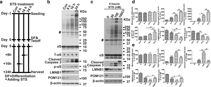
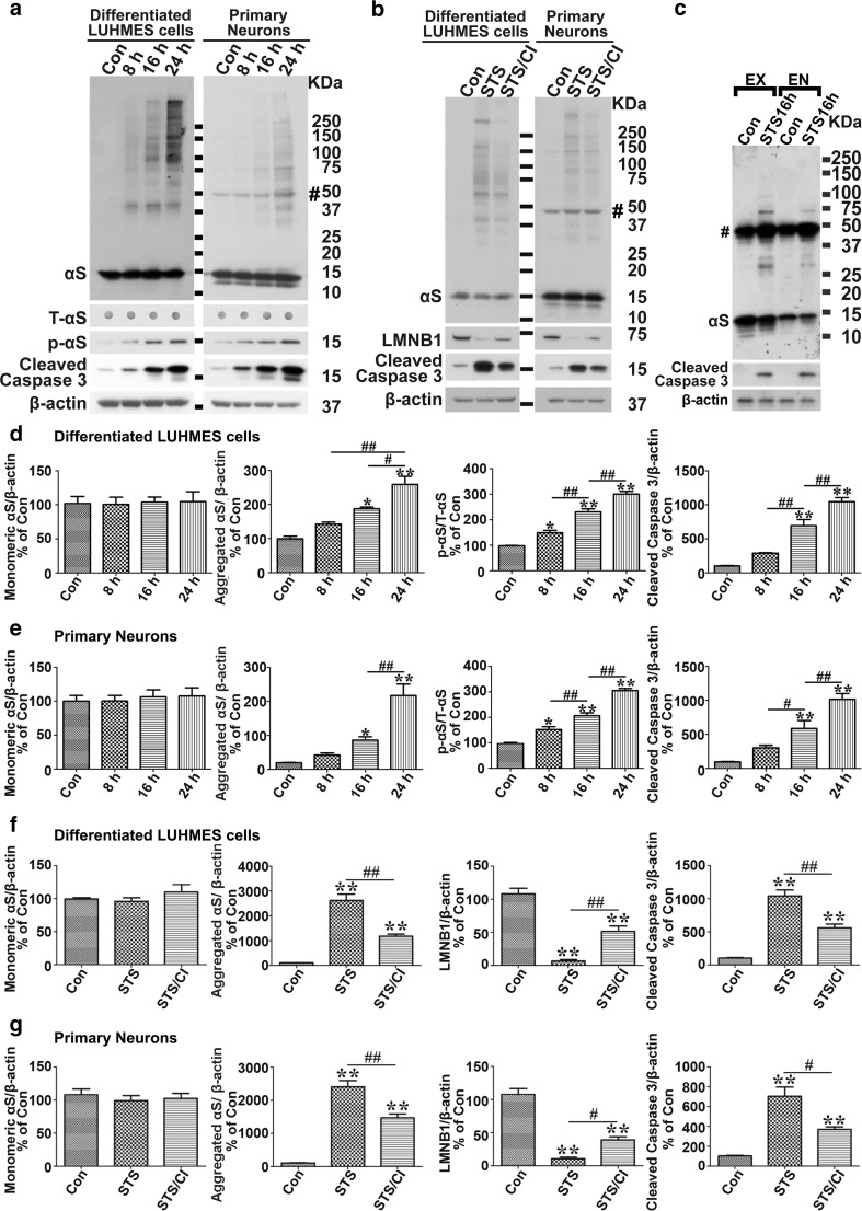
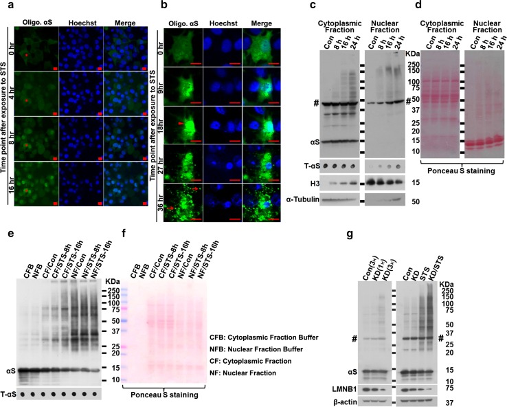
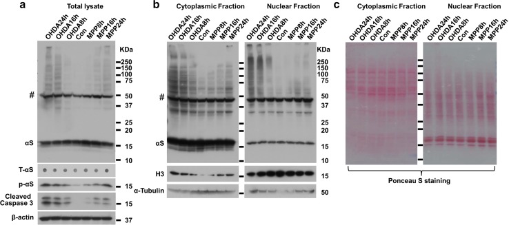
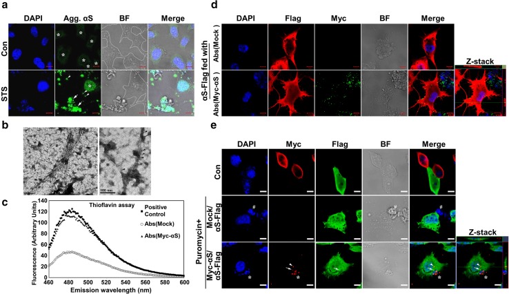
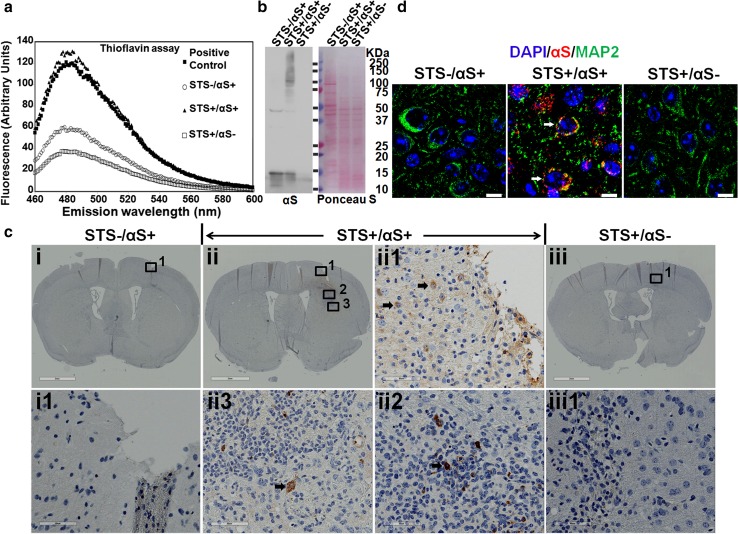
Cell-to-cell transmission of α-synuclein (αS) aggregates has been proposed to be responsible for progressive αS pathology in Parkinson disease (PD) and related disorders, including dementia with Lewy bodies. In support of this concept, a growing body of in vitro and in vivo experimental evidence shows that exogenously introduced αS aggregates can spread into surrounding cells and trigger PD-like pathology. It remains to be determined what factor(s) lead to initiation of αS aggregation that is capable of seeding subsequent propagation. In this study we demonstrate that filamentous αS aggregates form in neurons in response to apoptosis induced by staurosporine or other toxins-6-hydroxy-dopamine and 1-methyl-4-phenylpyridinium (MPP+). Interaction between αS and proaggregant nuclear factor(s) is associated with disruption of nuclear envelope integrity. Knocking down a key nuclear envelop constituent protein, lamin B1, enhances αS aggregation. Moreover, in vitro and in vivo experimental models demonstrate that aggregates released upon cell breakdown can be taken up by surrounding cells. Accordingly, we suggest that at least some αS aggregation might be related to neuronal apoptosis or loss of nuclear membrane integrity, exposing cytosolic α-synuclein to proaggregant nuclear factors. These findings provide new clues to the pathogenesis of PD and related disorders that can lead to novel treatments of these disorders. Specifically, finding ways to limit the effects of apoptosis on αS aggregation, deposition, local uptake and subsequent propagation might significantly impact progression of disease.

Keywords: 6OHDA; Aggregation; Apoptosis; Lamin B1; MPP+; Nuclear membrane integrity; Parkinson’s disease; Proaggregant nuclear factors; α-Synuclein.


Figures

Fig. 1
Fig. 1
Apoptosis induces αS aggregation and phosphorylation in BE(2)-M17D/3D5 cells. a The basic paradigm for cell differentiation, αS induction and staurosporine (STS) treatment. BE(2)-M17D/3D5 cells with 5 days of differentiation and αS induction were exposed to 25 nM STS for 0, 8, 16 and 24 h before harvest. b STS treatment of BE(2)-M17D/3D5 cells induces apoptosis, αS aggregation and Serine 129 phosphorylation, and degradation of lamin B1 (LMNB1) and nuclear pore membrane protein 121 KDa (POM121) in a time-dependent manner. c High dose of STS treatment can rapidly trigger apoptosis and other aforementioned effects which can be significantly prevented by caspase inhibitor. BE(2)-M17D/3D5 cells with 5 days of differentiation and αS induction were exposed to 50 and 100 nM for 4 h before harvest. A group of 100 nM STS-treated sibling cultures was pretreated with caspase inhibitor (CI). All cell lysates were subjected to SDS-PAGE followed by western blotting with antibodies to interesting proteins. Dot blotting of cell lysates was used to detect total αS level (T-αS) for quantification of p-αS/T-αS immunoreactivities. Molecular weight standards were included as references. Arrow denotes a specific cleaved caspase 3 band in BE(2)-M17D/3D5 cells pretreated with caspase inhibitor. Number sign (#) denotes a non-specific Syn1-immunoreactive band on western blots of lysates from BE(2)-M17D/3D5 cells. d, e Statistical analysis of immunoreactivity of various proteins shown in (b and c). Bar graphs in d and e summarize the results of quantitative analyses of immunoreactivities of various proteins in cell lysates from three independent experiments represented by b and c with normalization against β-actin or T-αS immunoreactivities. The average values of control group (Con or 0 nM) were set as 100 %. Error bars represent standard error of the mean (*p < 0.05, **p < 0.01, comparing to control; #p < 0.05, ##p < 0.01, comparing subsets linked by line, n = 3)
Fig. 2
Fig. 2
Apoptosis induces αS aggregation and phosphorylation in differentiated LUHMES and primary neurons. a STS treatment induces apoptosis, αS aggregation and phosphorylation in a time-dependent manner in differentiated LUHMES cells and primary neurons. Differentiated LUHMES cells and primary neuronal cultures overexpressing αS via infection with lentivirus carrying αS were, respectively, exposed to 100 nM STS for 0, 8, 16 and 24 h before being harvested. b Caspase inhibitor (CI) prevents STS-induced apoptosis and αS aggregation in differentiated LUHMES cells and primary neuronal cultures. Cultures without STS treatment were used as control (Con). STS-treated sibling cultures were pretreated with caspase inhibitor (STS/CI) to inhibit apoptosis. c Apoptosis induces aggregation of endogenous αS in mouse primary cultures. Primary neuronal cultures with only endogenously expressed αS (EN) or with exogenously overexpression of αS (EX) via infection with lentivirus carrying αS were exposed to 100 nM STS or vehicle (Con) for 16 h before harvest. All cell lysates were subjected to SDS-PAGE followed by western blotting with antibodies to proteins of interesting. Dot blotting of cell lysates was used to detect the total αS level (T-αS) for quantification of p-αS/T-αS immunoreactivities. Molecular weight standards were included as references. Number sign (#) denotes a non-specific Syn1-immunoreactive band on western blots of lysates from primary cultures. dg Statistical analyses of immunoreactivities of various proteins shown in a and b. Bar graphs in dg summarize results of quantitative analyses of immunoreactivities of various proteins in cell lysates from three independent experiments represented by a left, a right, b left and b right with normalization against β-actin or T-αS immunoreactivities. The average values of control group (Con) were set as 100 %. Error bars represent standard error of the mean (*p < 0.05, **p < 0.01, comparing to control; #p < 0.05, ##p < 0.01, comparing subsets linked by line, n = 3)
Fig. 3
Fig. 3
Disruption of nuclear envelope plays an essential role in the rapid αS aggregation during neuronal apoptosis. a, b αS aggregation in response to apoptosis induction predominantly occurred in nuclei rather than cytoplasm in live H4/V1S-SV2 cells. H4/V1S-SV2 cells cultured in Nunc™ Lab-Tek chambered cover glass system were induced to express H4/V1S-SV2 for 4 days, then maintained in media containing STS and Hoechst 33342 and subjected to time-lapse imaging using confocal fluorescence microscopy. A low magnification of live cell imaging demonstrates that about 94 % cells (30–32) had oligomeric αS (Olig. αS) predominantly in nuclei of cells (see red asterisk * in a) under 16 h of STS treatment. High magnification of live imaging of a single cell showed that the majority of αS aggregates appears first in nuclei (arrow head in b at 18 h) and then distributed to cytoplasm before finally being released to surrounding cells due to cell breakdown (arrows in b at 36 h). Scar bar: 10 µm. c, d Fractionation of cytoplasmic and nuclear proteins revealed the dynamic distribution of αS aggregates and other proteins in apoptotic neurons. Differentiated BE(2)-M17D/3D5 cells with 5 days of αS induction were, respectively, exposed to 25 nM for 0, 8, 16 and 24 h before harvest. c Nuclear and cytoplasm fractions were isolated and subjected to SDS-PAGE followed by western blotting. Dot blots were used to detect total αS (T-αS) for evaluation of the dynamic changes of T-αS in different fractions. α-Tubulin and histone H3 were probed to confirm there was no contamination between cytoplasmic and nuclear fraction in non-treated control group. d Ponceau S staining was performed before immunostaining to demonstrate total protein in each group. e, f Proaggregant nuclear factor(s) diffuse into cytoplasm with progression of apoptosis. Nuclear and cytoplasm fractions from null M17D neuroblastoma cells with 0, 8 and 16 h of STS treatment were, respectively, isolated. Fresh recombinant αS (10 µg) was incubated with the same amount of protein from each fraction for 30 min at 37 °C. The same amount of recombinant αS incubated with only cytoplasmic fraction buffer (CFB) or nuclear fraction buffer (NFB) was used as control. e Samples were subjected to SDS-PAGE followed by western blotting with monoclonal antibody to αS (Syn1). Dot blotting was used to confirm the comparable level of input recombinant αS in each sample. f Ponceau S staining was performed to demonstrate comparable levels of protein in all samples of cytoplasmic and nuclear fractions. g Disintegration of nuclear envelope is associated with rapid αS aggregation in neuronal cells in response to apoptosis induction. Differentiated BE(2)-M17D/3D5 cells with 5 days of αS induction infected with lentivirus carrying shRNA of LMNB1 and control vector were exposed to media with or without 25 nM STS for 16 h. Lentivirus infection was performed on the day 3 of culture. Left: one arbitrary unit (1x) or 3 units (3x) of lentivirus were applied to cells to obtain different extent of LMNB1 knockdown (KD). For control (Con), 3 units (3x) of lentivirus was used. Right: one arbitrary unit of lentivirus was used for both control (Con) and LMNB1 knockdown (KD). Cell lysates were subjected to SDS-PAGE and western blotting. Number sign (#) denotes a non-specific Syn1-immunoreactive band on western blots of lysates from BE(2)-M17D/3D5 cells in (c, g)
Fig. 4
Fig. 4
Aggregation and phosphorylation of αS as well as the distribution of αS aggregates in neuronal cells are induced by neurotoxins MPP+ and 6OHDA. Differentiated BE(2)-M17D/3D5 cells with 5 days of αS induction were, respectively, exposed to MPP+ or 6-6OHDA for 8, 16 and 24 h before harvest. Cells without treatment were used as control (Con). Each group of cells was divided into two parts for extraction of total cell lysates or isolation of nuclear and cytoplasm fractions. Samples were subjected to SDS-PAGE followed by western blotting with antibodies to proteins of interest. Dot blots of cell lysates were used to detect the total αS level (T-αS). Molecular weight standards were included as references. a Analysis of total lysates showed that aggregation and Serine 129 phosphorylation of αS gradually increased with progression of apoptosis in neuronal cells treated with MPP+ or 6OHDA. b Fractionation of cytoplasmic and nuclear proteins revealed that dynamic changes in distribution of αS aggregates and other proteins in neuronal cells induced by MPP+ or 6-6OHDA treated were similar to those induced by STS. c. Ponceau S staining was performed to demonstrate that cytoplasmic and nuclear fraction have comparable levels of total proteins
Fig. 5
Fig. 5
Apoptotic bodies derived from neuronal cells with αS overexpression contain αS aggregates that are taken up by surrounding cells. a Apoptotic bodies with disrupted nuclear envelope have αS aggregates. Differentiated BE(2)-M17D/3D5 cells with 5 days of αS expression were induced to enter late apoptotic stage by STS treatment for 36 h and then subjected to immunocytochemical staining with LB509 antibody and nuclear counterstaining with DAPI. Cells with intact nuclei are outlined (white line) in bright-field (BF) microscopy image. Signals of aggregated αS (Agg. αS) in cells without STS treatment (Con) are enhanced to demonstrate the cell morphology since the laser setting used for imaging STS-treated cells was not visible in control cells. Arrows and arrowheads denote apoptotic bodies and other smaller structures. The asterisk (*) labeled the location of intact nuclei in both groups. b Ultrastructural evaluation of sarkosyl-insoluble fractions prepared from apoptotic bodies. Apoptotic bodies were isolated from differentiated BE(2)-M17D/3D5 cells with 5 days of αS expression and 36 h of STS treatment and then subjected to fractionation to obtain Sarkosyl-insoluble fraction. (Left) Sarkosyl-insoluble fraction adsorbed on EM grids and negatively stained with 5 % uranyl acetate revealed filamentous assemblies of diameter about 8–10 nm. (Right) The filaments were decorated with 5 nm gold particles (arrows) by immunogold labeling using an anti-αS antibody, thus verifying that they were assembled from αS. Similar filamentous structures were not detected in apoptotic bodies from null M17D cells (data not shown). Scale bar: 100 nm. c Thioflavin T binding assay for sonicated apoptotic bodies derived from differentiated Myc-αS cells and mock-transfected cells with 36 h of puromycin treatment to induce apoptosis. Apoptotic bodies containing αS-positive filaments shown in (b) were used as positive control. The PBS solution was used as blank. d Immunocytochemistry showed Myc immunoreactivity outside and inside αS-Flag cells treated with sonicated apoptotic bodies from Myc-αS cells. Similar findings are not observed in control cells treated with sonicated apoptotic bodies from null M17D cells. Z-stack imaging confirmed the internalization of Myc-positive αS aggregates. e Neuronal cells can take up αS aggregates released from surrounding apoptotic cells. αS-Flag cells were co-cultured with Myc-αS or mock transfected M17D cells at equal density and subjected to differentiation for 5 days and then maintained in media with or without puromycin for another 5 days. After that, cells were fixed for immunocytochemical staining with antibodies to Myc and Flag and counterstaining with DAPI. Arrows and arrowheads denote Myc-positive aggregates from apoptotic bodies. Asterisk (*) and number sign (#), respectively, marked the apoptotic bodies from Myc-αS or mock transfected cells. Scale bar: 10 µm for all pictures
Fig. 6
Fig. 6
Apoptotic bodies derived αS aggregates can be taken up by neuronal cells in vivo upon inoculation into mouse brain. Apoptotic bodies from STS-treated BE(2)-M17D/3D5 cells with or without αS induction, respectively, referred to as STS+/αS+ and STS+/αS−, were isolated and injected into brains of mice around age of 12 months. Cell lysates from BE(2)-M17D/3D5 cells with αS induction but without STS treatment was also included as another control (STS-/αS+). All injected materials were subjected to a thioflavin T assay to confirm the formation of filamentous αS and with SDS-PAGE/western blotting to confirm SDS-insolubility of αS aggregates. Apoptotic bodies containing αS-positive filaments shown in Fig. 5b were used as positive control for (a). Sections from the injected brain region were subjected to (c) immunohistochemical staining with LB509 to human αS and (d) immunofluorescence staining with LB509 and MAP2 to confirm that neuronal uptake of human αS aggregates can be only observed in mouse brain injected with apoptotic bodies (STS+/αS+). Black arrows in (c) denote neuron-like cells with internalized αS aggregates. White arrows in (d) denote MAP2 and human αS aggregates positive neurons. Scale bar: 2 mm for i, ii and iii, 60 µm for i1, ii1 to ii3 and iii1 in (c), 10 µm for (d)

References

    1. Cookson MR. The biochemistry of Parkinson’s disease. Ann Rev Biochem. 2005;74:29–52. doi: 10.1146/annurev.biochem.74.082803.133400. - DOI - PubMed
    1. Acquatella-Tran Van Ba I, Imberdis T, Perrier V. From prion diseases to prion-like propagation mechanisms of neurodegenerative diseases. Int J Cell Biol. 2013;2013:975832. doi: 10.1155/2013/975832. - DOI - PMC - PubMed
    1. Visanji NP, Brooks PL, Hazrati LN, Lang AE. The prion hypothesis in Parkinson’s disease: Braak to the future. Acta Neuropathol Comm. 2013;1:2. doi: 10.1186/2051-5960-1-2. - DOI - PMC - PubMed
    1. Braak H, Del Tredici K, Rub U, de Vos RA, Jansen Steur EN, Braak E. Staging of brain pathology related to sporadic Parkinson’s disease. Neurobiol Aging. 2003;24:197–211. doi: 10.1016/S0197-4580(02)00065-9. - DOI - PubMed
    1. Kordower JH, Chu Y, Hauser RA, Freeman TB, Olanow CW. Lewy body-like pathology in long-term embryonic nigral transplants in Parkinson’s disease. Nat Med. 2008;14:504–506. doi: 10.1038/nm1747. - DOI - PubMed

Publication types

MeSH terms