Review
. 2016 Apr 21;45(8):2075-89.
doi: 10.1039/c5cs00693g. Epub 2016 Feb 3.

Stimulated Raman scattering microscopy: an emerging tool for drug discovery

Affiliations
Review

Stimulated Raman scattering microscopy: an emerging tool for drug discovery

W J Tipping et al. Chem Soc Rev. .

Abstract

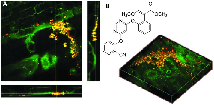
Optical microscopy techniques have emerged as a cornerstone of biomedical research, capable of probing the cellular functions of a vast range of substrates, whilst being minimally invasive to the cells or tissues of interest. Incorporating biological imaging into the early stages of the drug discovery process can provide invaluable information about drug activity within complex disease models. Spontaneous Raman spectroscopy has been widely used as a platform for the study of cells and their components based on chemical composition; but slow acquisition rates, poor resolution and a lack of sensitivity have hampered further development. A new generation of stimulated Raman techniques is emerging which allows the imaging of cells, tissues and organisms at faster acquisition speeds, and with greater resolution and sensitivity than previously possible. This review focuses on the development of stimulated Raman scattering (SRS), and covers the use of bioorthogonal tags to enhance sample detection, and recent applications of both spontaneous Raman and SRS as novel imaging platforms to facilitate the drug discovery process.


Figures

Fig. 1
Fig. 1. Electromagnetic radiation interacting with a vibrating molecule. When incident radiation (ω 0) interacts with a chemical species, it can be elastically scattered (Rayleigh scattering) or inelastically scattered (Raman scattering) by an amount, ω m which corresponds to the energy of a molecular transition in the molecule. In the case of Raman scattering, the scattered photon may have a lower energy compared with the incoming photon (Stokes scattering, ω 0ω m), alternatively the scattered photon may have a higher energy than the incident photon (anti-Stokes scattering, ω 0 + ω m).
Fig. 2
Fig. 2. Schematic energy level diagrams for spontaneous Raman, stimulated Raman scattering (SRS) and coherent anti-Stokes Raman scattering (CARS) processes. (A) In spontaneous Raman, the pump beam, ω p, is directed on to the sample generating a red-shifted signal ω s, due to inelastic scattering. (B) During SRS two laser beams at frequencies ω p and ω S are incident upon the sample, such that when the frequency difference (Δω = ω pω S) matches a molecular vibration in the sample (Ω), stimulated emission occurs. (C) CARS microscopy is a complex advanced Raman imaging technique, involving a four-beam mixing process probing at the anti-Stokes frequency (ω aS).
Fig. 3
Fig. 3. Label-free imaging of SKBr3 cells by Raman microscopy. (A) Spontaneous Raman spectrum of SKBr3 cell pellet measured using 785 nm excitation. Key bands are indicted, with wavenumbers measured in cm–1. (B) Spontaneous Raman image of two fixed SKBr3 cells, in the region 2100–3100 cm–1, with principal component analysis to identify different regions of the cell. Blue: principal component 1, yellow: principal component 2, red: principal component 3, black: principal component 4. Scale bar 10 μm. (C) Molecular distribution maps of fixed SKBr3 cells by Raman microscopy, (i) extracted image at 1033 cm–1 (C–H in-plane of phenylalanine); (ii) extracted image at 1657 cm–1 (CO, amide-I); (iii) extracted image at 2880 cm–1 (CH2, lipid); and (iv) extracted image at 2939 cm–1 (CH3). Scale bars 5 μm.
Fig. 4
Fig. 4. Output spectrum associated with SRS process. The Stokes beam experiences an intensity gain resulting from the stimulated excitation of a Raman active band within the target molecule (SRG). Conversely, the pump beam experiences an intensity loss during this process (SRL).
Fig. 5
Fig. 5. A schematic representation of a SRS microscope. A pump beam and an intensity-modulated Stokes beam are both temporally and spatially synchronized before being focused onto cells. When the energy difference between the pump photons and the Stokes photons matches the molecular vibration (Ω) of a bond, those particular bonds are efficiently driven from their vibrational ground state to their vibrational excited state, passing through a virtual state. For each excited bond, a photon in the pump beam is annihilated (Raman loss) and a photon in the Stokes beam is created (Raman gain). These intensity changes in the pump beam are extracted using a lock-in amplifier and can provide a quantitative map of the targeted vibrating chemical bonds.
Fig. 6
Fig. 6. Label free microscopy of squamous cell carcinoma cells by stimulated Raman scattering microscopy. (A) Stimulated Raman scattering image of fixed SCC cells, (i) acquired at 2953 cm–1 (CH3); (ii) acquired at 2850 cm–1 (CH2, lipid); (iii) acquired at 1655 cm–1 (CO, amide-1); and (iv) acquired at 1700 cm–1 (cellular silent region). Scale bars 5 μm.
Fig. 7
Fig. 7. Structures of molecules containing alkyne groups, which act as bioorthogonal markers for spontaneous Raman and SRS imaging. Erlotinib is a clinical drug which has an inherent alkyne bond, EdU is a thymidine analogue modified with an alkyne bond, Hpg is an alkyne containing methionine analogue, ALK-16 is a palmitic acid analogue modified with an alkyne group, 3-OPG is a glucose analogue modified with a propargyl group and Ac4ManNAI is an alkyne-containing glycan analogue.
Fig. 8
Fig. 8. Strategies for intracellular visualisation of biomolecules. (A) Bioorthogonal ligation strategy illustrated using a CuAAC click reaction. (B) Bioorthogonal Raman-active tagging strategy. A small Raman reporter installed onto, or inherent to, the biomolecule of choice is used as a marker to perform direct spontaneous Raman or SRS imaging.
Fig. 9
Fig. 9. Live SRS imaging of de novo synthesis of DNA. (A) Live HeLa cells incubated with EdU alone (100 μM) alkyne on; and with hydroxyurea (10 mM) control. (B) Time-lapse images of a dividing cell incubated with EdU (100 μM). Scale bars: 10 μm. Adapted from ref. 60 with permission. Copyright (2014) Nature Publishing group.
Fig. 10
Fig. 10. Spontaneous Raman imaging of erlotinib. (A) Representative spontaneous Raman spectrum of erlotinib showing clearly the strong alkyne band at 2110 cm–1. Spectrum acquired at 785 nm excitation. (B) Spontaneous Raman imaging of SW480 cells treated with erlotinib (∼100 μM, 12 h) (i) spontaneous Raman image reconstructed from the C–H stretching intensity; (ii) spontaneous Raman image constructed from the CC stretching intensity; (iii) overlay of panels (i) and (ii) showing the drug is clustered at the EGFR protein at the cell membrane. Adapted from ref. 49 with permission. Copyright (2014) Royal Society of Chemistry publishing.
Fig. 11
Fig. 11. Recent examples of biomolecules studied by SRS and spontaneous Raman imaging. (A) Rhabduscin. (B) SRS microscopy analysing the spatial localisation of rhabduscin based on the vibrational resonance of the isonitrile functional group at 2121 cm–1. (i) Rhabduscin is localised at the periphery of E. coli cells heterologously expressing the rhabduscin pathway; (ii) the background signal is low in the cells lacking the rhabduscin gene cluster. Scale bar 10 μm. Adapted from ref. 51 with permission from the National Academy of Sciences. (C) FCCP (D) amygadalin (E) mestranol and its conjugation to an inorganic coordination complex via a CuAAC click reaction.
Fig. 12
Fig. 12. Incorporation of Raman-active isotopologues into proteins and biomolecules. (A) Metabolic incorporation of labelled amino acids in auxotrophic cell lines. (B) Raman-active metabolic precursors: deuteriated phenylalanine, 15N-labelled ammonium chloride, 13C-labelled glucose, 13C-labelled phenylalanine.
Fig. 13
Fig. 13. Development of a three colour palette of alkyne tags. (A) Structures of unlabelled, mono, and bis 13C-labelled 5-ethynyl-2′-deoxyuridine (EdU). (B) Spontaneous Raman spectra of HeLa cells incubated with the three isotopically edited EdU's (0.1 mM, 15 h). Spectra were acquired from the nuclear region of fixed cells following incubation with either 1, 2 or 3. The spectra were normalised at the alkyne peak, and are displayed from 2000–2170 cm–1. (C) Live cell SRS imaging of DNA synthesis in HeLa cells incubated with isotopically edited EdUs. For each sample incubated with either 1, 2, or 3. Images are acquired in 5 different Raman channels: 1655 cm–1 (amide I band), 2000 cm–1 (off-resonant), 2048 cm–1 (on-resonant with 3), 2077 cm–1 (on-resonant with 2), and 2125 cm–1 (on-resonant with 1) in sequential mode. Images are acquired in 512 × 512 pixels with a pixel dwell time of 40 μs. Image adapted from ref. 81 with permission. Copyright (2014) American Chemical Society.
Fig. 14
Fig. 14. Structures of drugs which have been imaged by spontaneous Raman spectroscopy, and SRS microscopy, highlighting the key bonds and Raman stretching frequencies which have enabled their intracellular quantitation. Note: for clofazimine a range of peaks were used in the region 1100–1600 cm–1.
Fig. 15
Fig. 15. Maximum-intensity projection of 3D SRS images at 1305 cm–1 of BaF3/BCR-ABL1 cells incubated with (A) imatinib (20 μM, 4 h), (B) nilotinib (20 μM, 4 h), and (C) DMSO only. Scale bar 5 μM. Adapted from ref. 86 with permission. Copyright (2014) Nature Publishing Group.
Fig. 16
Fig. 16. Compositional analysis of intracellular compartments in whole C. elegans worms by hsSRS imaging, k-means clustering, and MCR analysis. (A and B) MCR-retrieved concentration maps of neutral fat droplets, lysosome-related organelles (LROs), oxidised lipids, and protein in the body of whole wild type worms (A) and daf-2 mutants (B). Scale bar: 50 μm. (C) Zoom-in of intestine cells indicated in (B). (D–G) MCR-reconstructed concentration images of LROs, fat droplets, oxidised lipids, and protein, respectively. Adapted from ref. 97 with permission. Copyright (2014) Wiley-VCH Verlag GmbH & Co kGaA, Weinheim.
Fig. 17
Fig. 17. SRS imaging of mouse ear skin with topically applied terbinafine hydrochloride (TH). (A) Terbinafine hydrochloride. (B) SRS imaging at the viable epidermis layer (z = 20 μm), (C) SRS imaging at the sebaceous gland layer (z = 40 μm) of mouse ear skin. In both (B and C), composite images show both protein (1655 cm–1) and lipid (2845 cm–1) distributions, and the alkyne images indicate that the TH penetrates via the lipidic phase. Scale bars, 20 μm. Adapted from ref. 60 with permission. Copyright (2014) Nature Publishing Group.
Fig. 18
Fig. 18. Application of azoxystrobin to maize leaves. (A) Azoxystrobin applied to a maize leaf (red = 2225 cm–1 from the CN bond, green = 2930 cm–1 from the CH3 vibrations) to show crystalline deposits of the chemical on the leaf surface, which appear yellow/orange due to contrast from the CH bonds (green) and CN bonds (red) which have been combined. (B) Chemical structure of azoxystrobin. Adapted from ref. 112 with permission. Copyright (2013) American Chemical Society.
None
W. J. Tipping
None
M. Lee
None
A. Serrels
None
V. G. Brunton
None
A. N. Hulme

References

    1. Kola I., Landis J. Nat. Rev. Drug Discovery. 2004;3:711–716. - PubMed
    1. Paul S. M., Mytelka D. S., Dunwiddie C. T., Persinger C. C., Munos B. H., Lindborg S. R., Schacht A. L. Nat. Rev. Drug Discovery. 2010;9:203–214. - PubMed
    1. Ocana A., Pandiella A., Siu L. L., Tannock I. F. Nat. Rev. Clin. Oncol. 2011;8:200–209. - PubMed
    1. Swinney D. C., Anthony J. Nat. Rev. Drug Discovery. 2011;10:507–519. - PubMed
    1. Moreno L., Pearson A. D. Expert Opin. Drug Discovery. 2013;8:363–368. - PubMed

Publication types

MeSH terms

LinkOut - more resources