Review
. 2016:2016:1384658.
doi: 10.1155/2016/1384658. Epub 2015 Dec 29.

Nanoparticles-Assisted Stem Cell Therapy for Ischemic Heart Disease

Affiliations
Review

Nanoparticles-Assisted Stem Cell Therapy for Ischemic Heart Disease

Kai Zhu et al. Stem Cells Int. 2016.

Abstract

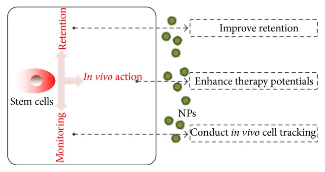
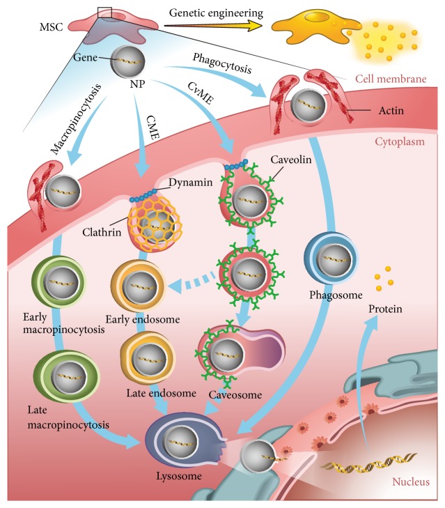
Stem cell therapy has attracted increasing attention as a promising treatment strategy for cardiac repair in ischemic heart disease. Nanoparticles (NPs), with their superior physical and chemical properties, have been widely utilized to assist stem cell therapy. With the help of NPs, stem cells can be genetically engineered for enhanced paracrine profile. To further understand the fate and behaviors of stem cells in ischemic myocardium, imaging NPs can label stem cells and be tracked in vivo under multiple modalities. Besides that, NPs can also be used to enhance stem cell retention in myocardium. These facts have raised efforts on the development of more intelligent and multifunctional NPs for cellular application. Herein, an overview of the applications of NPs-assisted stem cell therapy is given. Key issues and future prospects are also critically addressed.


Figures

Figure 1
Figure 1
Schematic illustration of NPs-assisted stem cell therapy.
Figure 2
Figure 2
Schematic illustrations of potential mechanisms of NPs-based endocytosis while delivering therapeutic gene into MSCs.
Figure 3
Figure 3
In vivo magnetic resonance images of mouse hearts injected with superparamagnetic NPs-loaded, cardiac-differentiated mouse embryonic stem cells. Scans were performed at 1, 7, and 28 days after cell transplantation in both the long-axis and short-axis orientations. NPs-loaded cells were shown as dark regions (white arrows) in the left ventricular wall in or near the infarct zone. Reprinted with permission from Ebert et al. [61].

Similar articles

Cited by

References

    1. Go A. S., Mozaffarian D., Roger V. L., et al. Heart disease and stroke statistics—2013 update: a report from the American Heart Association. Circulation. 2013;127(1):e6–e245. doi: 10.1161/cir.0b013e31828124ad. - DOI - PMC - PubMed
    1. Blecker S., Paul M., Taksler G., Ogedegbe G., Katz S. Heart failure-associated hospitalizations in the United States. Journal of the American College of Cardiology. 2013;61(12):1259–1267. doi: 10.1016/j.jacc.2012.12.038. - DOI - PMC - PubMed
    1. Ptaszek L. M., Mansour M., Ruskin J. N., Chien K. R. Towards regenerative therapy for cardiac disease. The Lancet. 2012;379(9819):933–942. doi: 10.1016/S0140-6736(12)60075-0. - DOI - PubMed
    1. Deedwania P. C., Carbajal E. V. Medical therapy versus myocardial revascularization in chronic coronary syndrome and stable angina. The American Journal of Medicine. 2011;124(8):681–688. doi: 10.1016/j.amjmed.2011.02.036. - DOI - PubMed
    1. Deb S., Wijeysundera H. C., Ko D. T., Tsubota H., Hill S., Fremes S. E. Coronary artery bypass graft surgery vs percutaneous interventions in coronary revascularization: a systematic review. Journal of the American Medical Association. 2013;310(19):2086–2095. doi: 10.1001/jama.2013.281718. - DOI - PubMed

LinkOut - more resources