

Posttraumatic Sphingomonas paucimobilis Endophthalmitis
- PMID: 26839724
- PMCID: PMC4709607
- DOI: 10.1155/2015/192864
Posttraumatic Sphingomonas paucimobilis Endophthalmitis
Abstract
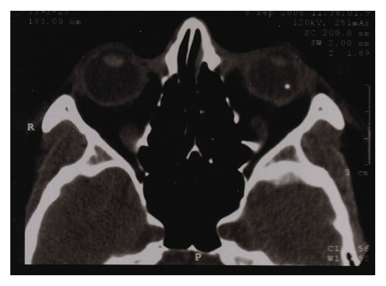
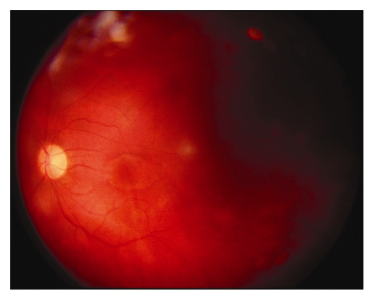
A rare case of Sphingomonas paucimobilis endophthalmitis secondary to a penetrating globe injury with a retained intraocular foreign body is presented. A 30-year-old man presented with severe pain following a penetrating left eye injury. Visual acuity (VA) was 6/120. Slit-lamp examination revealed perforation of the temporal cornea and iris, hypopyon, and a fibrinous membrane covering the pupil. Ultrasonography showed dense vitreous infiltration and an orbital CT-scan confirmed the presence of a metallic foreign body in the vitreous cavity. Topical and systemic therapy were initiated. Pars-plana vitrectomy combined with phacoemulsification was performed in order to remove the foreign body; vitreous samples were acquired and Sphingomonas paucimobilis, sensitive to ceftazidime, was identified. To the best of our knowledge, this is the first report of Sphingomonas paucimobilis endophthalmitis following penetrating ocular injury. In this case, Sphingomonas paucimobilis was not resistant to antibiotics. This allowed for a good healing response following vitrectomy despite the fact that long-term retinal complications resulted in low VA.
Figures
References
-
- Miyazaki Y., Oka S., Yamaguchi S., Mizuno S., Yano I. Stimulation of phagocytosis and phagosome-lysosome fusion by glycosphingolipids from Sphingomonas paucimobilis . Journal of Biochemistry. 1995;118(2):271–277. - PubMed
-
- Boken D. J., Romero J. R., Cavalieri S. J. Sphingomonas paucimobilis bacteremia: four cases and review of the literature. Infectious Diseases in Clinical Practice. 1998;7(6):286–291. doi: 10.1097/00019048-199808000-00011. - DOI
LinkOut - more resources
Full Text Sources
Other Literature Sources
Miscellaneous
