. 2016 Jan 28:4:e1628.
doi: 10.7717/peerj.1628. eCollection 2016.

Protective effects of the extracts of Barringtonia racemosa shoots against oxidative damage in HepG2 cells

Affiliations

Protective effects of the extracts of Barringtonia racemosa shoots against oxidative damage in HepG2 cells

Kin Weng Kong et al. PeerJ. .

Abstract

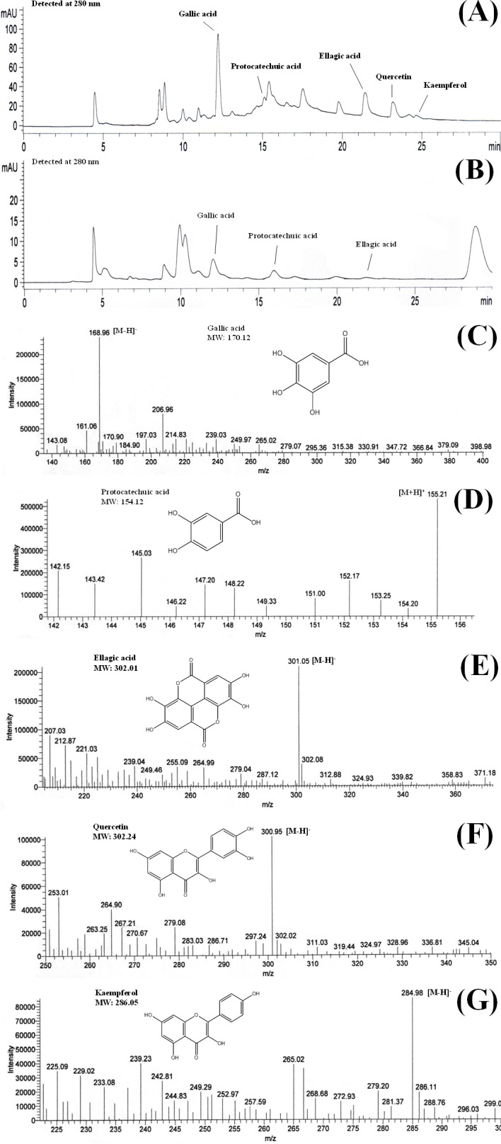
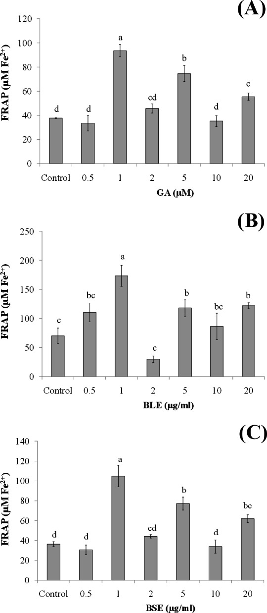
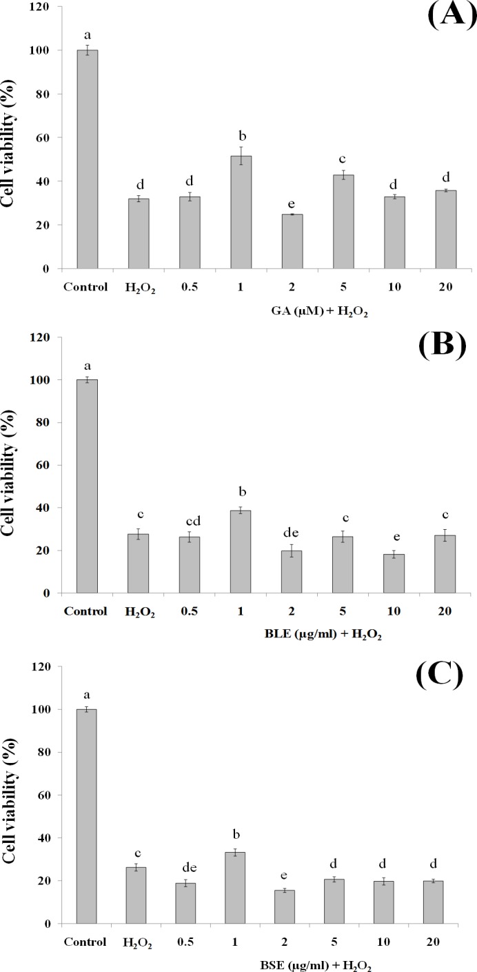
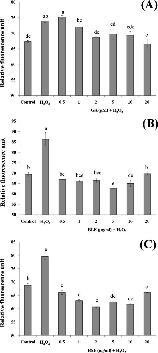
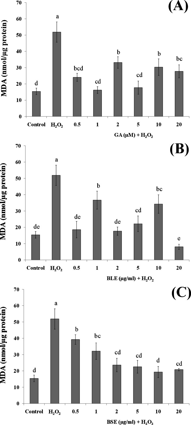
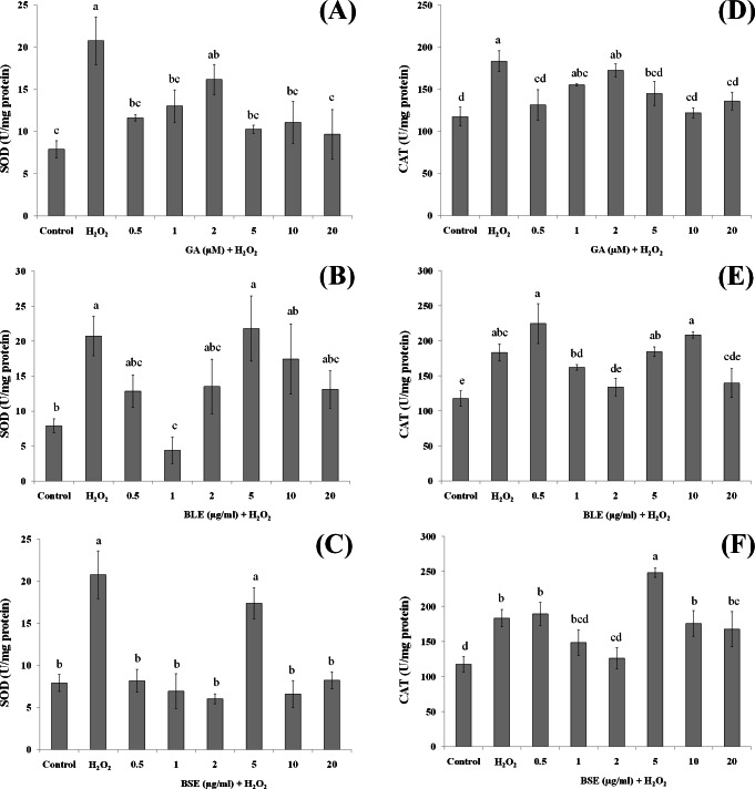
Barringtonia racemosa is a tropical plant with medicinal values. In this study, the ability of the water extracts of the leaf (BLE) and stem (BSE) from the shoots to protect HepG2 cells against oxidative damage was studied. Five major polyphenolic compounds consisting of gallic acid, ellagic acid, protocatechuic acid, quercetin and kaempferol were identified using HPLC-DAD and ESI-MS. Cell viability assay revealed that BLE and BSE were non-cytotoxic (cell viabilities >80%) at concentration less than 250 µg/ml and 500 µg/ml, respectively. BLE and BSE improved cellular antioxidant status measured by FRAP assay and protected HepG2 cells against H2O2-induced cytotoxicity. The extracts also inhibited lipid peroxidation in HepG2 cells as well as the production of reactive oxygen species. BLE and BSE could also suppress the activities of superoxide dismutase and catalase during oxidative stress. The shoots of B. racemosa can be an alternative bioactive ingredient in the prevention of oxidative damage.

Keywords: Antioxidant enzymes; Barringtonia racemosa; HPLC-ESI-MS; Lipid peroxidation; Oxidative stress; Polyphenols.


Conflict of interest statement

The authors declare there are no competing interests.

Figures

Figure 1
Figure 1. Chromatograms of (A) BLE and (B) BSE and the mass to charge ratio (mz) of (C) gallic acid, (D) protocatechuic acid, (E) ellagic acid, (F) quercetin and (G) kaempferol.
The chromatograms were obtained from HPLC-DAD analyses while the mz was obtained from ESI-MS analyses. [M + H]+ and [M − H] are the ions of the detected compounds obtained from the negative and positive full scan modes. BLE, Leaf water extract of B. racemosa; BSE, Stem water extract of B. racemosa.
Figure 2
Figure 2. The effects of (A) gallic acid, (B) BLE and (C) BSE on antioxidant status of HepG2 cells.
Cells (5 × 10³ cells/well) were treated with gallic acid, BLE and BSE for 24 h and cellular antioxidant status was measured using FRAP assay. Results are expressed as means ± SEM. Values with different letters are significantly different at p < 0.05. BLE, Leaf water extract of B. racemosa; BSE, Stem water extract of B. racemosa; GA, Gallic acid; Control, untreated cells.
Figure 3
Figure 3. The cytoprotective effects of (A) gallic acid, (B) BLE and (C) BSE on HepG2 cells following H2O2-induced oxidative damage.
Cells (5 × 10³ cells/well) were pre-treated with gallic acid, BLE and BSE for 24 h prior to H2O2-induced oxidative damage. Results are expressed as means ± SEM. Values with different letters are significantly different at p < 0.05. BLE, Leaf water extract of B. racemosa; BSE, Stem water extract of B. racemosa; GA, Gallic acid.
Figure 4
Figure 4. The effects of (A) gallic acid, (B) BLE and (C) BSE on ROS production of HepG2 cells following H2O2-induced oxidative damage.
Cells (5 × 10³ cells/well) were pre-treated with the plant extracts or gallic acid for 24 h prior to H2O2-induced oxidation. ROS production was determined by measuring relative fluorescence, using DCFH-DA probe. Values with different letters are significantly different at p < 0.05. BLE, Leaf water extract of B. racemosa; BSE, Stem water extract of B. racemosa; GA, Gallic acid; Control, negative control; H2O2, positive control.
Figure 5
Figure 5. The effects of (A) gallic acid, (B) BLE and (C) BSE on lipid peroxidation of HepG2 cells following H2O2-induced oxidative damage.
Cells (1.5 × 10⁵ cells/well) were pre-treated with the plant extracts or gallic acid for 24 h prior to H2O2-induced oxidation. MDA was measured by the TBARS method. Values with different letters are significantly different at p < 0.05. BLE, Leaf water extract of B. racemosa; BSE, Stem water extract of B. racemosa; GA: Gallic acid; Control: negative control; H2O2, positive control.
Figure 6
Figure 6. The effects of gallic acid, BLE and BSE on activities of SOD (A–C) and CAT (D–F) in HepG2 cells following H2O2-induced oxidative damage.
Cells (1.5 × 10⁵ cells/well) were pre-treated with the plant extracts or gallic acid for 24 h prior to induction of oxidation with H2O2. Results are expressed as means ± SEM. Values with different letters are significantly different at p < 0.05. BLE, Leaf water extract of B. racemosa; BSE, Stem water extract of B. racemosa; GA, Gallic acid.

Similar articles

Cited by

References

    1. Alía M, Mateos R, Ramos S, Lecumberri E, Bravo L, Goya L. Influence of quercetin and rutin on growth and antioxidant defense system of a human hepatoma cell line (HepG2) European Journal of Nutrition. 2006a;45:19–28. doi: 10.1007/s00394-005-0558-7. - DOI - PubMed
    1. Alía M, Ramos S, Mateos R, Granado-Serrano AB, Bravo L, Goya L. Quercetin protects human hepatoma HepG2 against oxidative stress induced by tert-butyl hydroperoxide. Toxicology and Applied Pharmacology. 2006b;212:110–118. doi: 10.1016/j.taap.2005.07.014. - DOI - PubMed
    1. Benzie IFF, Strain JJ. The ferric reducing ability of plasma (FRAP) as a measure of ‘antioxidant power’: the FRAP assay. Analytical Biochemistry. 1996;239:70–76. doi: 10.1006/abio.1996.0292. - DOI - PubMed
    1. Boulton DW, Walle UK, Walle T. Extensive binding of the bioflavonoid quercetin to human plasma proteins. Journal of Pharmacy and Pharmacology. 1998;50:243–249. - PubMed
    1. Bradford MM. A rapid and sensitive method for the quantitation of microgram quantities of protein utilizing the principle of protein dye binding. Analytical Biochemistry. 1976;72:248–254. doi: 10.1016/0003-2697(76)90527-3. - DOI - PubMed