. 2016:2016:8178936.
doi: 10.1155/2016/8178936. Epub 2015 Dec 29.

The Actions of Lyophilized Apple Peel on the Electrical Activity and Organization of the Ventricular Syncytium of the Hearts of Diabetic Rats

Affiliations

The Actions of Lyophilized Apple Peel on the Electrical Activity and Organization of the Ventricular Syncytium of the Hearts of Diabetic Rats

Elideth Martínez-Ladrón de Guevara et al. J Diabetes Res. 2016.

Abstract

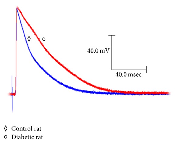
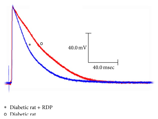
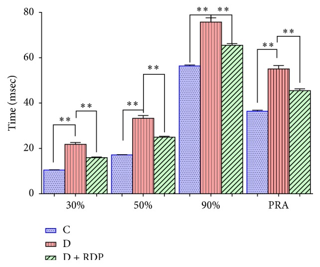
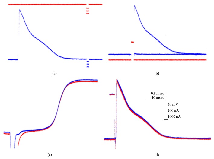
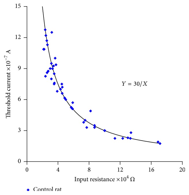
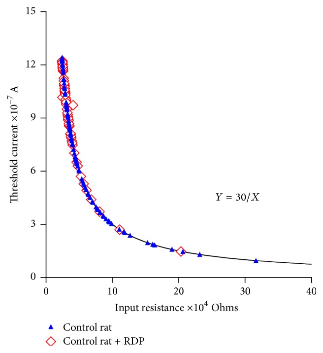
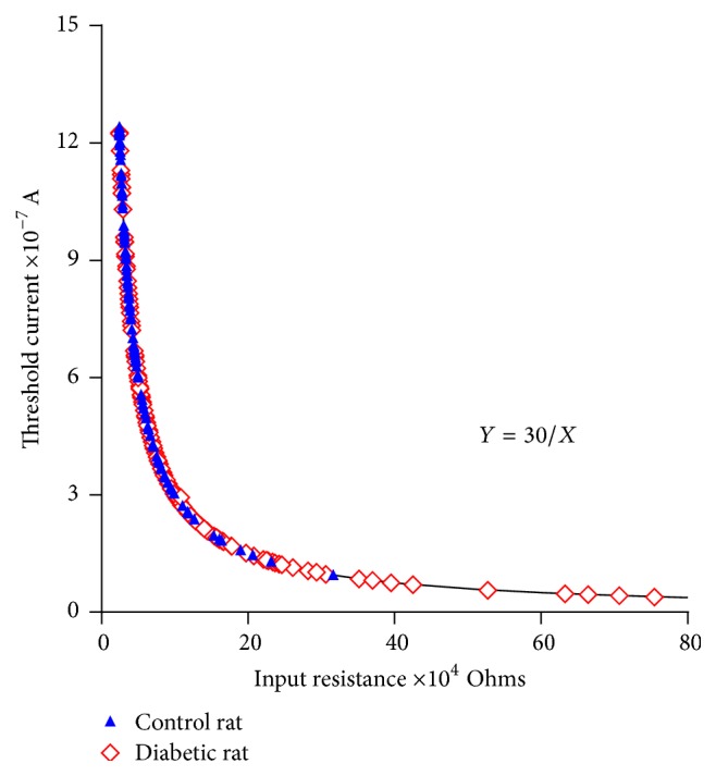
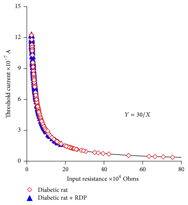
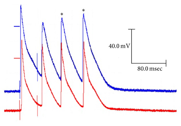
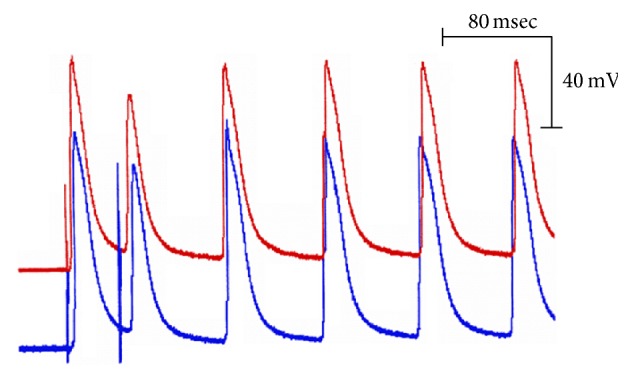
This study was designed to examine the effects of lyophilized red delicious apple peel (RDP) on the action potentials (APs) and the input resistance-threshold current relationship. The experiments were performed on isolated papillary heart muscles from healthy male rats, healthy male rats treated with RDP, diabetic male rats, and diabetic male rats treated with RDP. The preparation was superfused with oxygenated Tyrode's solution at 37°C. The stimulation and the recording of the APs, the input resistance, and the threshold current were made using conventional electrophysiological methods. The RDP presented no significant effect in normal rats. Equivalent doses in diabetic rats reduced the APD and ARP. The relationship between input resistance and threshold current established an inverse correlation. The results indicate the following: (1) The functional structure of the cardiac ventricular syncytium in healthy rats is heterogeneous, in terms of input resistance and threshold current. Diabetes further accentuates the heterogeneity. (2) As a consequence, conduction block occurs and increases the possibility of reentrant arrhythmias. (3) These modifications in the ventricular syncytium, coupled with the increase in the ARP, are the adequate substrate so that, with diabetes, the heart becomes more arrhythmogenic. (4) RDP decreases the APD, the ARP, and most syncytium irregularity caused by diabetes.


Figures

Figure 1
Figure 1
Changes in the action potential with diabetes. Representative action potentials obtained from a cell of the papillary muscle of the left ventricle of the heart of control rats and diabetic rats at a basic cycle of 500 milliseconds.
Figure 2
Figure 2
Action of the apple peel on the action potentials of diabetic rats. Typical transmembrane potentials obtained in the papillary muscles of the heart of diabetic rats and diabetic rats with apple peel at a basic cycle of 500 milliseconds.
Figure 3
Figure 3
Mean values ± SEM of the quantified parameters of the action potentials recorded in the papillary muscles of the hearts of control rats (n = 225), diabetic rats (n = 110), and diabetic rats receiving apple peel for 90 days (n = 169) at a basic cycle of 500 milliseconds. The symbols (∗∗) indicate that the differences between the different groups are significant (p < 0.05).
Figure 4
Figure 4
Procedure for determining the input resistance. Steps followed to quantify the input resistance and the intracellular thresholds in the papillary muscles of the hearts of the control rats: (a) injection of the pulse current in late diastole to evaluate the input resistance; (b) determination of the intracellular threshold; (c) simultaneity and shape of the depolarization phase of the action potentials; (d) zero potential of both records obtained from the same cell.
Figure 5
Figure 5
Fit relationship of the experimental input resistance-current threshold values (blue) with respect to the theoretical curve (black). Values obtained in the cells of the papillary muscle of the heart of the control rats. The continuous curve corresponds to an equilateral hyperbola defined by the equation Y = K/X, where K = 30 mV. For each input resistance value, there is a corresponding threshold current value adequately conforming to the theoretical curve, which is established by the statistical analysis of R 2 = 0.92 and p < 0.05.
Figure 6
Figure 6
Input resistance-current threshold relationship in control rats and control rats with RDP treatment (150 mg/kg). The values of the input resistance were calculated using the equation of the equilateral hyperbola, Y = K/X, where Y is the threshold current quantified experimentally and K = 30 mV is the constant value. Filled symbols: control rats; unfilled symbols: control rats + RDP.
Figure 7
Figure 7
Input resistance versus threshold current relationship in the control rats and diabetic rats. The relationship obtained in the papillary muscle of the heart of the control rats and the rats with diabetes. The continuous line corresponds to the theoretical equilateral hyperbola, Y = 30/X. Filled symbols: control rats; unfilled symbols: diabetic rats.
Figure 8
Figure 8
Changes in the input resistance-current threshold relationships in diabetic rats and diabetic rats treated with RDP; 150 mg/kg for 90 days. Filled symbols: diabetic rats; unfilled symbols: diabetic + RDP.
Figure 9
Figure 9
Reentrant activity initiated by the application of an early extrasystole. The traces shown correspond to action potentials recorded in the papillary muscles of diabetic rats, in the area proximal (upper trace) and the area distal (lower trace) to the stimulation site, at a basic cycle of 500 milliseconds. Observe that, after the implementation of the extrasystole, two responses (asterisks) not evoked by stimulation appear, corresponding to a type of reentrant activity.
Figure 10
Figure 10
Change of direction in the propagation of the wavefront. Observe that the first three responses (basic, test, and first reentrant) are propagated from the stimulation site towards the rest of the preparation, whereas the last three following reentrant responses propagate in the opposite direction.

Similar articles

References

    1. McGhie T. K., Hudault S., Lunken R. C. M., Christeller J. T. Apple peels, from seven cultivars, have lipase-inhibitory activity and contain numerous ursenoic acids as identified by LC-ESI-QTOF-HRMS. Journal of Agricultural and Food Chemistry. 2012;60(1):482–491. doi: 10.1021/jf203970j. - DOI - PubMed
    1. Francini A., Sebastiani L. Phenolic compounds in apple (Malus x domestica Borkh.): compounds characterization and stability during postharvest and after processing. Antioxidants. 2013;2(3):181–193. doi: 10.3390/antiox2030181. - DOI - PMC - PubMed
    1. Hyson D. A. A comprehensive review of apples and apple components and their relationship to human health. Advances in Nutrition. 2011;2(5):408–420. doi: 10.3945/an.111.000513. - DOI - PMC - PubMed
    1. Andre C. M., Greenwood J. M., Walker E. G., et al. Anti-inflammatory procyanidins and triterpenes in 109 apple varieties. Journal of Agricultural and Food Chemistry. 2012;60(42):10546–10554. doi: 10.1021/jf302809k. - DOI - PubMed
    1. Bizjak J., Mikulic-Petkovsek M., Stampar F., Veberic R. Changes in primary metabolites and polyphenols in the peel of ‘braeburn’ apples (Malus domestica Borkh.) during advanced maturation. Journal of Agricultural and Food Chemistry. 2013;61(43):10283–10292. doi: 10.1021/jf403064p. - DOI - PubMed

Publication types

LinkOut - more resources