. 2016 Feb 3;11(2):e0142476.
doi: 10.1371/journal.pone.0142476. eCollection 2016.

Therapeutic Benefits of Induced Pluripotent Stem Cells in Monocrotaline-Induced Pulmonary Arterial Hypertension

Affiliations

Therapeutic Benefits of Induced Pluripotent Stem Cells in Monocrotaline-Induced Pulmonary Arterial Hypertension

Wei-Chun Huang et al. PLoS One. .

Abstract

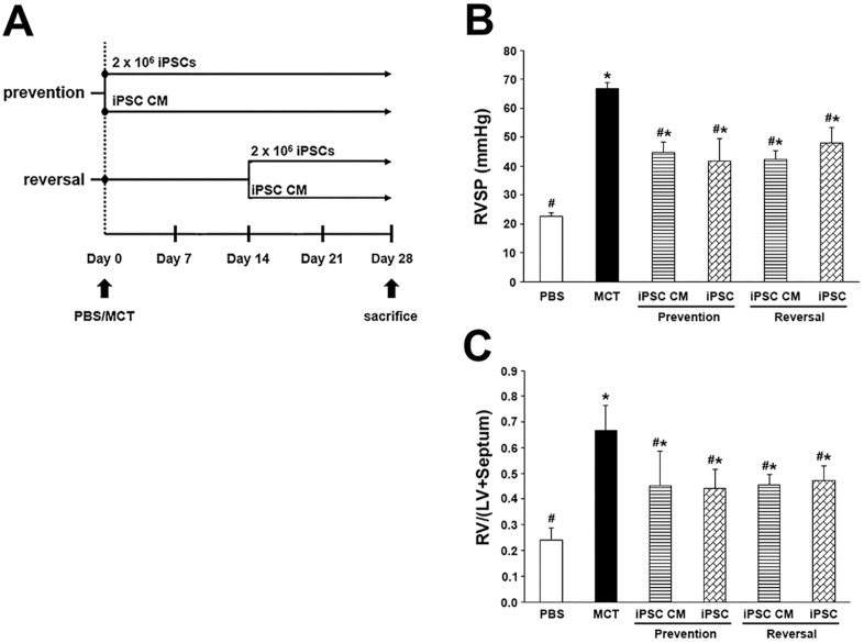
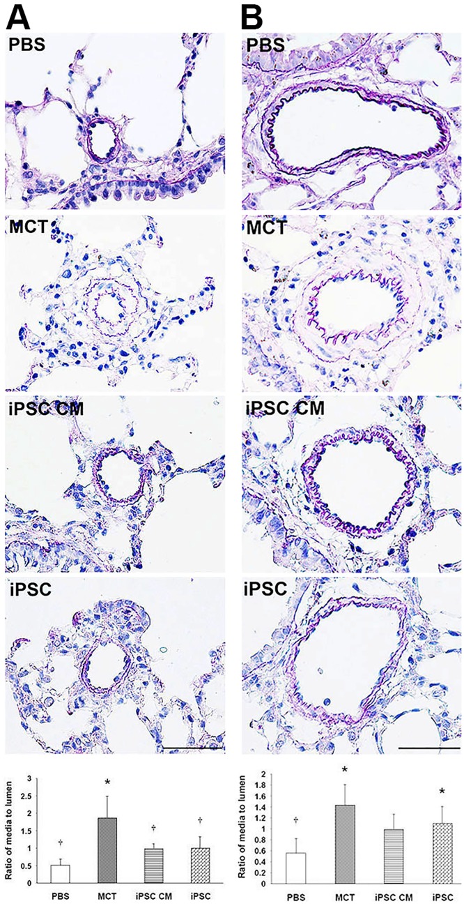
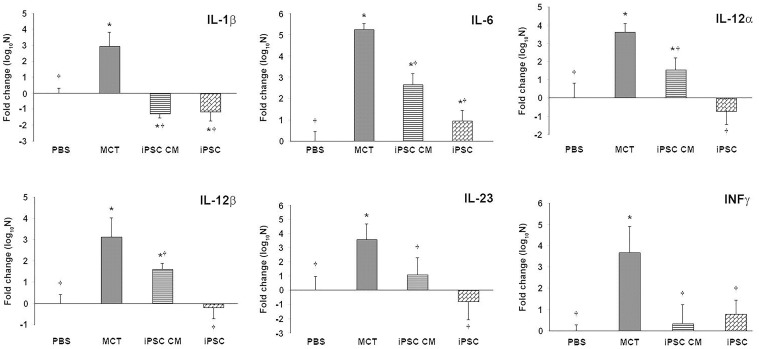
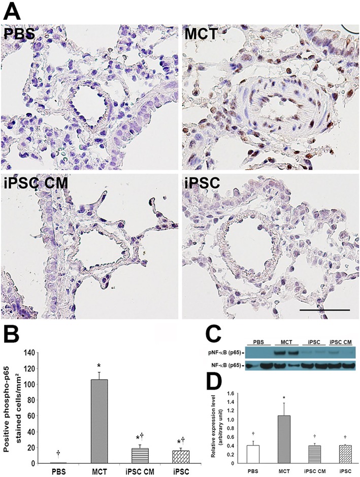
Pulmonary arterial hypertension (PAH) is characterized by progressive increases in vascular resistance and the remodeling of pulmonary arteries. The accumulation of inflammatory cells in the lung and elevated levels of inflammatory cytokines in the bloodstream suggest that inflammation may play a role in PAH. In this study, the benefits of induced pluripotent stem cells (iPSCs) and iPSC-conditioned medium (iPSC CM) were explored in monocrotaline (MCT)-induced PAH rats. We demonstrated that both iPSCs and iPSC CM significantly reduced the right ventricular systolic pressure and ameliorated the hypertrophy of the right ventricle in MCT-induced PAH rats in models of both disease prevention and disease reversal. In the prevention of MCT-induced PAH, iPSC-based therapy led to the decreased accumulation of inflammatory cells and down-regulated the expression of the IL-1β, IL-6, IL-12α, IL-12β, IL-23 and IFNγ genes in lung specimens, which implied that iPSC-based therapy may be involved in the regulation of inflammation. NF-κB signaling is essential to the inflammatory cascade, which is activated via the phosphorylation of the NF-κB molecule. Using the chemical inhibitor specifically blocked the phosphorylation of NF-κB, and in vitro assays of cultured human M1 macrophages implied that the anti-inflammation effect of iPSC-based therapy may contribute to the disturbance of NF-κB activation. Here, we showed that iPSC-based therapy could restore the hemodynamic function of right ventricle with benefits for preventing the ongoing inflammation in the lungs of MCT-induced PAH rats by regulating NF-κB phosphorylation.


Conflict of interest statement

Competing Interests: The authors have declared that no competing interests exist.

Figures

Fig 1
Fig 1. Characterization of murine induced pluripotent stem cells.
Murine iPSCs were plated onto mitomycin c-treated MEFs for 3 days and formed cobblestone-like colonies. (B) iPSCs demonstrated specific and strong alkaline phosphatase activity. Scale bar = 100 μm. (C) Immunocytochemistry revealed the expression of iPSC marker SSEA-1. Scale bar = 100 μm. (D) Gel electrophoresis demonstrates the semi-quantitative RT-PCR results for several key genes unique to iPSCs. Nat1 served as an internal control.
Fig 2
Fig 2. Effect of iPSC-based therapy on RVSP and the weight ratio of RV/(LV+Septum).
Representative scheme of iPSC-based therapy on MCT-induced PAH. (B) Hemodynamic changes in RVSP were analyzed across animal groups at day 28 prior to sacrifice. (C) RV hypertrophy was measured by comparing the weight ratio of RV/(LV+Septum) of different groups. The results are shown as the mean ± SD for 6 rats of each group. *p<0.05 vs. PBS group; #p<0.05 vs. MCT group.
Fig 3
Fig 3. EVG analysis of pulmonary artery hypertrophy.
The progressive narrowing and thickening of blood vessels (particularly beginning in arterioles) is typical in PAH. We performed EVG staining on paraffin-embedded lung sections. Blood vessels were divided into two classes according to their diameter: (A) < 100 μm and (B) > 100 μm. The thickness of blood vessels was evaluated by calculating the ratio of the area of the media layer to that of the lumen. Our findings were most dramatic in blood vessels under 100 μm in diameter; iPSCs treatments significantly prevented the progressive thickening of the arterioles. The area ratio of the media to the lumen was represented as the means ± SD, and the value for each group was averaged over 10 vascular images taken from different fields. Scale bar = 100 μm. *p<0.05 vs. PBS group; p<0.05 vs. MCT group.
Fig 4
Fig 4. iPSC-based therapy attenuated inflammation in the lung tissue of MCT-induced PAH rats.
Inflammation in the lungs of MCT-induced PAH rats was analyzed via immunohistochemistry of inflammatory markers in paraffin-embedded sections. (A) Positive staining for macrophage marker CD68. (B) Quantification of CD68 stain calculated as the number of positive cells per sq. millimeter from 10 separate images of different fields; data are represented as the means ± SD. (D-E) The same protocol was used to detect MHC-II; the results were confirmed via western blotting (C and F, CD68 and MHC-II, respectively). iPSC-based therapy attenuated the level of inflammation in MCT-induced PAH rats. Scale bar = 100 μm. *p<0.05 vs. PBS group; p<0.05 vs. MCT group.
Fig 5
Fig 5. iPSC-based therapy downregulated the expression of proinflammatory genes in the lungs of MCT-induced PAH rats.
Semi-quantitative RT-PCR (qRT-PCR) was used to detect the expression of several proinflammatory genes including IL-1β, IL-6, IL-12α, IL-12β, IL-23 and INFγ. RNA extracted from the lung tissue of the MCT-induced PAH rats and controls was reverse transcribed for semi-quantitative RT-PCR. The data were normalized according to 36b4 mRNA levels and represented as a value relative to the PBS control group. The results are shown as the means ± SD for 5–8 rats per group. *p<0.05 vs. PBS group; p<0.05 vs. MCT group.
Fig 6
Fig 6. iPSC-based treatment suppressed inflammation in polarized classical human macrophages.
The effects of murine iPSCs on inflammation were evaluated in the context of co-culture with polarized classical human macrophages. Primitive human macrophages were differentiated from PBMCs and subsequently polarized (via the stimulation of LPS and IFNγ) to become proinflammatory M1 macrophages. To examine paracrine effects of iPSCs in vitro, cells were separated using a permeable membrane (A), thereby mimicking the conditioned media treatment in rats. (B) Secretion of the inflammatory cytokines TNFα and IL-1β was measured via ELISA. Data were accumulated from 3–5 independent assays. (C) The immunocytochemical analysis of inflammatory markers CD68 and MHC-II was performed on co-cultured iPSCs and M1 macrophages. CD68 and MHC-II (Texas Red) and iPSC-specific marker SSEA-1 (FITC) are shown. Nuclei were counterstained with Hoechst 33342. (D) Genes specifically expressed in M1 macrophages were compared across groups of iPSC-based treatments using semi-quantitative RT-PCR. The results are represented as the means ± SD; values were normalized according to 36b4 mRNA levels and represented as the fold change relative to the PBS control group. *p<0.05 vs. PBS group; p<0.05 vs. MCT group. Scale bar = 10 μm.
Fig 7
Fig 7. iPSC-based therapy regulated inflammation in MCT-induced PAH via the suppression of NF-κB phosphorylation.
Phosphorylation of NF-κB protein was detected via immunohistochemistry with a monoclonal antibody specific to the phosphorylated form of the NF-κB p65 subunit. (C) The expression level of phosphorylated NF-κB protein relative to the total was confirmed via western blot. (B and D) Quantification of immunohistochemistry and western blot, respectively. Both iPSC treatments demonstrated significant inhibitory effects on NF-κB phosphorylation compared with MCT treatment. The phosphorylated NF-κB stain was quantified as the number of positive cells per sq. millimeter from 10 separate images of different fields; results are given as the means ± SD. *p<0.05 vs. PBS group; p<0.05 vs. MCT group. Scale bar = 100 μm.

References

    1. Humbert M, Morrell NW, Archer SL, Stenmark KR, MacLean MR, Lang IM, et al. Cellular and molecular pathobiology of pulmonary arterial hypertension. J Am Coll Cardiol. 2004;43 (12 Suppl S): 13S–24S. - PubMed
    1. Budhiraja R, Tuder RM, Hassoun PM. Endothelial dysfunction in pulmonary hypertension. Circulation. 2004;109(2): 159–65. - PubMed
    1. Smith JD, Trogan E, Ginsberg M, Grigaux C, Tian J, Miyata M, et al. Decreased atherosclerosis in mice deficient in both macrophage colony-stimulating factor (op) and apolipoprotein E. Proc Natl Acad Sci U S A. 1995;92(18): 8264–8268. - PMC - PubMed
    1. Zhao YY, Zhao YD, Mirza MK, Huang JH, Potula HH, Vogel SM, et al. Persistent eNOS activation secondary to caveolin-1 deficiency induces pulmonary hypertension in mice and humans through PKG nitration. J Clin Invest. 2009;119(7): 2009–2018. 10.1172/JCI33338 - DOI - PMC - PubMed
    1. Gomez-Arroyo JG, Farkas L, Alhussaini AA, Farkas D, Kraskauskas D, Voelkel NF, et al. The monocrotaline model of pulmonary hypertension in perspective. Am J Physiol Lung Cell Mol Physiol. 2012;302(4): L363–9. 10.1152/ajplung.00212.2011 - DOI - PubMed

Publication types

MeSH terms